. 2019 Jan 9:9:791.
doi: 10.3389/fendo.2018.00791. eCollection 2018.

Characterization of Non-hormone Expressing Endocrine Cells in Fetal and Infant Human Pancreas

Affiliations

Characterization of Non-hormone Expressing Endocrine Cells in Fetal and Infant Human Pancreas

Abu Saleh Md Moin et al. Front Endocrinol (Lausanne). .

Abstract

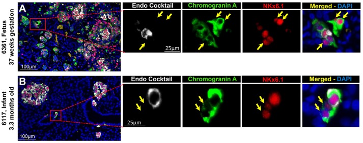
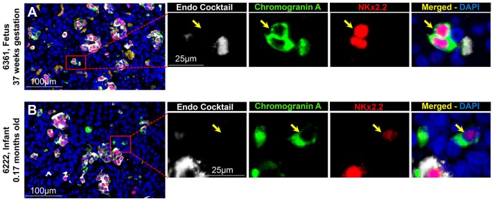
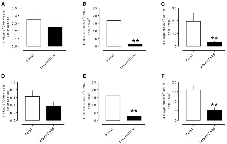
Context: Previously, we identified chromograninA positive hormone-negative (CPHN) cells in high frequency in human fetal and neonatal pancreas, likely representing nascent endocrine precursor cells. Here, we characterize the putative endocrine fate and replicative status of these newly formed cells. Objective: To establish the replicative frequency and transcriptional identity of CPHN cells, extending our observation on CPHN cell frequency to a larger cohort of fetal and infant pancreas. Design, Setting, and Participants: 8 fetal, 19 infant autopsy pancreata were evaluated for CPHN cell frequency; 12 fetal, 24 infant/child pancreata were evaluated for CPHN replication and identity. Results: CPHN cell frequency decreased 84% (islets) and 42% (clusters) from fetal to infant life. Unlike the beta-cells at this stage, CPHN cells were rarely observed to replicate (0.2 ± 0.1 vs. 4.7 ± 1.0%, CPHN vs. islet hormone positive cell replication, p < 0.001), indicated by the lack of Ki67 expression in CPHN cells whether located in the islets or in small clusters, and with no detectable difference between fetal and infant groups. While the majority of CPHN cells express (in overall compartments of pancreas) the pan-endocrine transcription factor NKX2.2 and beta-cell specific NKX6.1 in comparable frequency in fetal and infant/child cases (81.9 ± 6.3 vs. 82.8 ± 3.8% NKX6.1+-CPHN cells of total CPHN cells, fetal vs. infant/child, p = 0.9; 88.0 ± 4.7 vs. 82.1 ± 5.3% NKX2.2+-CPHN cells of total CPHN cells, fetal vs. infant/child, p = 0.4), the frequency of clustered CPHN cells expressing NKX6.1 or NKX2.2 is lower in infant/child vs. fetal cases (1.2 ± 0.3 vs. 16.7 ± 4.7 clustered NKX6.1+-CPHN cells/mm2, infant/child vs. fetal, p < 0.01; 2.7 ± 1.0 vs. 16.0 ± 4.0 clustered NKX2.2+-CPHN cells/mm2, infant/child vs. fetal, p < 0.01). Conclusions: The frequency of CPHN cells declines steeply from fetal to infant life, presumably as they differentiate to hormone-expressing cells. CPHN cells represent a non-replicative pool of endocrine precursor cells, a proportion of which are likely fated to become beta-cells. Precis : CPHN cell frequency declines steeply from fetal to infant life, as they mature to hormone expression. CPHN cells represent a non-replicative pool of endocrine precursor cells, a proportion of which are likely fated to become beta-cells.

Keywords: beta-cell; development; endocrine cells; maturation; replication; transcription factor.


Figures

Figure 1
Figure 1
The frequency of chromograninA positive hormone-negative (CPHN) cells decreases with age. Representative pancreatic sections from fetal (A) and infant-child (B–D) cases immunostained for Endocrine cocktail (insulin, glucagon, somatostatin, pancreatic polypeptide, and ghrelin) (white), chromograninA (green), and DAPI (blue). Yellow arrows show CPHN cells. Frequency of CPHN cells in islets (E) and clusters (F). Scale bars, 50 μm.
Figure 2
Figure 2
ChromograninA positive hormone-negative (CPHN) cells do not replicate during fetal and infant life. Representative pancreatic sections from fetal (A) and infant (B) donors immunostained for Endocrine cocktail (insulin, glucagon, somatostatin, pancreatic polypeptide, and ghrelin) (white), chromograninA (green), Ki67 (red), and DAPI (blue). Yellow arrows indicate CPHN cells. CPHN cells were rarely positive for Ki67 staining in both fetal and infant groups; no detectable difference was found in the frequency of replicative CPHN cells between fetal and infant pancreatic sections. Scale bars: 100 μm for low power and 50 μm for high magnification images.
Figure 3
Figure 3
ChromograninA positive hormone-negative (CPHN) cells express the endocrine differentiation transcription factor NKX6.1 in both fetal and infant pancreas. Representative pancreatic sections from fetal (A) and infant (B) donors immunostained for Endocrine cocktail (insulin, glucagon, somatostatin, pancreatic polypeptide, and ghrelin) (white), chromograninA (green), the transcription factor NKX6.1 (red) and DAPI (blue). Yellow arrows indicate CPHN cells. Scale bars: 100 μm for low power and 25 μm for high magnification images.
Figure 4
Figure 4
ChromograninA positive hormone-negative (CPHN) cells express the beta-cell differentiation transcription factor NKX2.2 in both fetuses and infants. Representative pancreatic sections from fetal (A) and infant (B) donors immunostained for Endocrine cocktail (insulin, glucagon, somatostatin, pancreatic polypeptide, and ghrelin) (white), chromograninA (green), the transcription factor NKX2.2 (red) and DAPI (blue). Yellow arrows indicate CPHN cells. Scale bars: 100 μm for low power and 25 μm for high magnification images.
Figure 5
Figure 5
Comparison of number of CPHN cells expressing transcription factors NKX6.1 and NKX2.2 in different compartments of fetal and infant/child pancreas. CPHN cells that express either NKX6.1 or NKX2.2 showed variability in their numbers among different compartments of the pancreas sections of both fetal and infant cases. The number of CPHN cells located within the islets that expressed either NKX6.1 (A) or NKX2.2 (D) was similar in fetal and infant/child cases but NKX6.1+ and NKX2.2+ CPHN cells found as clusters (B,E) or single cells (C,F) were significantly higher in fetal cases compared to infant/child cases. **p < 0.01, n = 7 for fetal cases and n = 10 for infant/child cases.

References

    1. Meier JJ, Kohler CU, Alkhatib B, Sergi C, Junker T, Klein HH, et al. . Beta-cell development and turnover during prenatal life in humans. Eur J Endocrinol. (2010) 162:559–68. 10.1530/EJE-09-1053 - DOI - PubMed
    1. Meier JJ, Butler AE, Saisho Y, Monchamp T, Galasso R, Bhushan A, et al. . Beta-cell replication is the primary mechanism subserving the postnatal expansion of beta-cell mass in humans. Diabetes (2008) 57:1584–94. 10.2337/db07-1369 - DOI - PMC - PubMed
    1. Kassem SA, Ariel I, Thornton PS, Scheimberg I, Glaser B. Beta-cell proliferation and apoptosis in the developing normal human pancreas and in hyperinsulinism of infancy. Diabetes (2000) 49:1325–33. 10.2337/diabetes.49.8.1325 - DOI - PubMed
    1. Butler AE, Dhawan S, Hoang J, Cory M, Zeng K, Fritsch H, et al. . beta-cell deficit in obese type 2 diabetes, a minor role of beta-cell dedifferentiation and degranulation. J Clin Endocrinol Metab. (2016) 101:523–32. 10.1210/jc.2015-3566 - DOI - PMC - PubMed
    1. Md Moin AS, Dhawan S, Shieh C, Butler PC, Cory M, Butler AE. Increased hormone-negative endocrine cells in the pancreas in type 1 diabetes. J Clin Endocrinol Metab. (2016) 101:3487–96. 10.1210/jc.2016-1350 - DOI - PMC - PubMed