

Blood Coagulation Following an Acute Ischemic Stroke
- PMID: 30687528
- PMCID: PMC6320462
- DOI: 10.12865/CHSJ.44.02.04
Blood Coagulation Following an Acute Ischemic Stroke
Abstract
Objective: Hemostasis is a complex physiological process that stops bleeding at the site of a vascular injury. Although the majority of vascular accidents are ischemic, the role of hypercoagulable state and stroke needs further investigation.
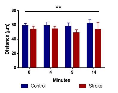
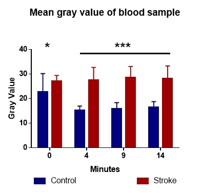
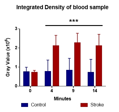
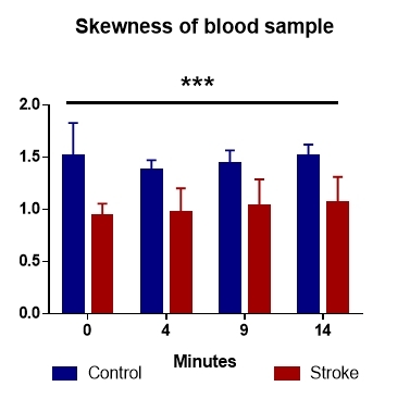
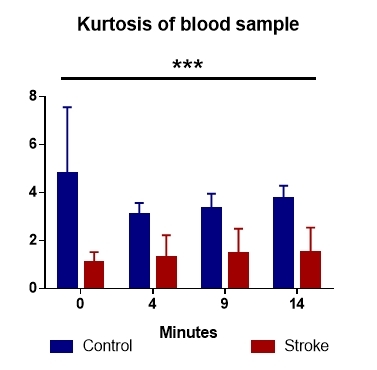
Materials and methods: Fresh whole blood was taken from 61 acute ischemic stroke patients and compared to 18 healthy subjects and investigated with optical coherence tomography imaging after initiating coagulation. We used an OCT1300SS system (Thorlabs) and did 3D scans. We then processed the images with ImageJ. For each image mean, integrated density, skewness and kurtosis of gray values were analyzed.
Results: Mean gray value and integrated intensity of sampled data showed an intrinsic difference detected with OCT. This difference was further confirmed by the data distribution analysis.
Conclusions: Results suggest, that normal blood coagulation, is not a random reaction while in the case of stroke patients, the relatively symmetrical distribution of gray values brings coagulation closer randomized process.
Keywords: cerebral ischemia; coagulation; optical coherence tomography.
Figures
Similar articles
-
Comparison of image sensitivity between conventional tensor-based and fast diffusion kurtosis imaging protocols in a rodent model of acute ischemic stroke.NMR Biomed. 2016 May;29(5):625-30. doi: 10.1002/nbm.3506. Epub 2016 Feb 26. NMR Biomed. 2016. PMID: 26918411 Free PMC article.
-
Trial design and reporting standards for intra-arterial cerebral thrombolysis for acute ischemic stroke.Stroke. 2003 Aug;34(8):e109-37. doi: 10.1161/01.STR.0000082721.62796.09. Epub 2003 Jul 17. Stroke. 2003. PMID: 12869717
-
Monitoring Acute Stroke Progression: Multi-Parametric OCT Imaging of Cortical Perfusion, Flow, and Tissue Scattering in a Mouse Model of Permanent Focal Ischemia.IEEE Trans Med Imaging. 2019 Jun;38(6):1427-1437. doi: 10.1109/TMI.2019.2895779. Epub 2019 Jan 31. IEEE Trans Med Imaging. 2019. PMID: 30714910 Free PMC article.
-
Prognostic Hemostasis Biomarkers in Acute Ischemic Stroke.Arterioscler Thromb Vasc Biol. 2019 Mar;39(3):360-372. doi: 10.1161/ATVBAHA.118.312102. Arterioscler Thromb Vasc Biol. 2019. PMID: 30700129 Free PMC article.
-
Hematologic disorders associated with ischemic stroke.J Neurol Sci. 1996 Sep 1;140(1-2):1-11. doi: 10.1016/0022-510x(96)00051-2. J Neurol Sci. 1996. PMID: 8866421 Review.
Cited by
-
Pulmonary Embolism in Acute Ischaemic Stroke: Evolving Evidence, Diagnostic Challenges, and a Novel Thromboinflammatory Axis Hypothesis.Int J Mol Sci. 2025 Jul 14;26(14):6733. doi: 10.3390/ijms26146733. Int J Mol Sci. 2025. PMID: 40724982 Free PMC article. Review.
-
The interaction effect of transfusion history and previous stroke history on the risk of venous thromboembolism in stroke patients: a prospective cohort study.Thromb J. 2023 Apr 17;21(1):41. doi: 10.1186/s12959-023-00487-2. Thromb J. 2023. PMID: 37069620 Free PMC article.
-
A novel cascade allows Metarhizium robertsii to distinguish cuticle and hemocoel microenvironments during infection of insects.PLoS Biol. 2021 Aug 4;19(8):e3001360. doi: 10.1371/journal.pbio.3001360. eCollection 2021 Aug. PLoS Biol. 2021. PMID: 34347783 Free PMC article.
-
Ivermectin's neuroprotective effects decrease with prolonged use after cerebral ischemia/reperfusion.Metab Brain Dis. 2025 Jun 25;40(6):234. doi: 10.1007/s11011-025-01657-z. Metab Brain Dis. 2025. PMID: 40560490
-
Optical coherence tomography microscopy in experimental traumatic brain injury.Microsc Res Tech. 2021 Mar;84(3):422-431. doi: 10.1002/jemt.23599. Epub 2020 Oct 3. Microsc Res Tech. 2021. PMID: 33009699 Free PMC article.
References
-
- Tanaka KA, Key NS, Levy JH. Blood coagulation: hemostasis and thrombin regulation. Anesthesia and analgesia. 2009;108(5):1433–1446. - PubMed
-
- de Lau, Leebeek FW, de Maat, Koudstaal PJ, Dippel DW. Screening for coagulation disorders in patients with ischemic stroke. Expert review of neurotherapeutics. 2010;10(8):1321–1329. - PubMed
LinkOut - more resources
Full Text Sources