

A Diagnostic Challenge: Pancreatic Cancer or Autoimmune Pancreatitis?
- PMID: 30687529
- PMCID: PMC6320463
- DOI: 10.12865/CHSJ.44.02.15
A Diagnostic Challenge: Pancreatic Cancer or Autoimmune Pancreatitis?
Abstract
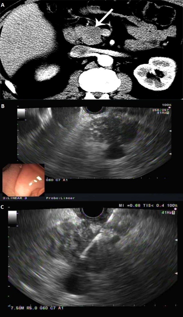
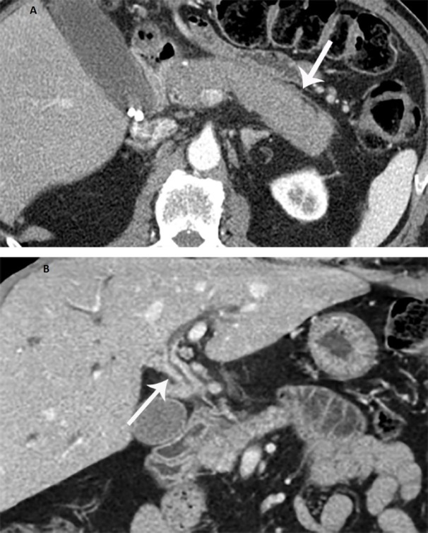
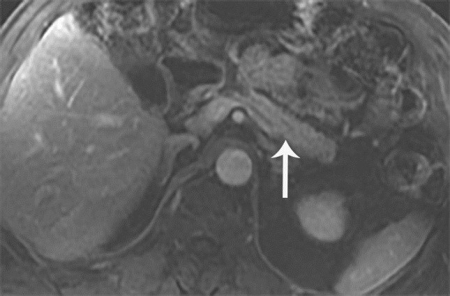
We report a rare case of seronegative autoimmune pancreatitis (AIP) that presented as a pancreatic focal lesion and was considered to be pancreatic cancer based on the clinical presentation and imaging findings. The endoscopic ultrasound-guided biopsies of the pancreatic mass revealed no malignant cells and the pancreatic swelling had become diffuse on repeat imaging. AIP was suspected and a trial of steroids was considered as a diagnostic and therapeutic method. The patient responded dramatically to corticosteroid treatment with resolution of symptoms and normal imagining and laboratory parameters. This case highlights the challenge in the diagnostic approach of a pancreatic mass.
Keywords: endoscopic ultrasound; endoscopic ultrasound-guided fine-needle aspiration; autoimmune pancreatitis; pancreatic cancer.
Figures
Similar articles
-
Autoimmune pancreatitis: role of endoscopy in diagnosis and treatment.Gastrointest Endosc Clin N Am. 2013 Oct;23(4):893-915. doi: 10.1016/j.giec.2013.06.005. Epub 2013 Jul 5. Gastrointest Endosc Clin N Am. 2013. PMID: 24079796 Review.
-
Pancreatic tuberculosis or autoimmune pancreatitis.Case Rep Med. 2014;2014:410142. doi: 10.1155/2014/410142. Epub 2014 Apr 15. Case Rep Med. 2014. PMID: 24839445 Free PMC article.
-
Branch Duct Intraductal Papillary Mucinous Neoplasms of the Pancreas Involving Type 1 Localized Autoimmune Pancreatitis with Normal Serum IgG4 Levels Successfully Diagnosed by Endoscopic Ultrasound-guided Fine-needle Aspiration and Treated without Pancreatic Surgery.Intern Med. 2017;56(10):1163-1167. doi: 10.2169/internalmedicine.56.8017. Epub 2017 May 15. Intern Med. 2017. PMID: 28502930 Free PMC article.
-
Comparison of endoscopic retrograde cholangiopancreatography with papillary biopsy and endoscopic ultrasound-guided pancreatic biopsy in the diagnosis of autoimmune pancreatitis.Pancreatology. 2015 May-Jun;15(3):259-64. doi: 10.1016/j.pan.2015.03.011. Epub 2015 Apr 1. Pancreatology. 2015. PMID: 25891790
-
A case of concurrent pancreatic intraepithelial neoplasia and type 1 autoimmune pancreatitis with marked pancreatic duct dilatation.Clin J Gastroenterol. 2016 Aug;9(4):266-71. doi: 10.1007/s12328-016-0666-3. Epub 2016 Jun 28. Clin J Gastroenterol. 2016. PMID: 27351197 Review.
References
-
- Klöppel G, Detlefsen S, Chari ST, Longnecker DS, Zamboni G. Autoimmune pancreatitis: the clinicopathological characteristics of the subtype with granulocytic epithelial lesions. Journal of gastroenterology. 2010;45(8):787–793. - PubMed
-
- Zhang L, Chari S, Smyrk TC, Deshpande V, Klöppel G, Kojima M, Liu X, Longnecker DS, Mino-Kenudson M, Notohara K. Autoimmune pancreatitis (AIP) type 1 and type 2: an international consensus study on histopathologic diagnostic criteria. Pancreas. 2011;40(8):1172–1179. - PubMed
-
- Hart PA, Zen Y, Chari ST. Reviews in basic and clinical gastroenterology and hepatology. Gastroenterology. 2015;149:39–51. - PubMed
-
- Sarles H, Sarles J-C, Muratore R, Guien C. Chronic inflammatory sclerosis of the pancreas—an autonomous pancreatic disease. The American journal of digestive diseases. 1961;6(7):688–698. - PubMed
LinkOut - more resources
Full Text Sources
Miscellaneous