

Mechanisms of cardiovascular disease in obstructive sleep apnoea
- PMID: 30687536
- PMCID: PMC6321896
- DOI: 10.21037/jtd.2018.08.56
Mechanisms of cardiovascular disease in obstructive sleep apnoea
Abstract
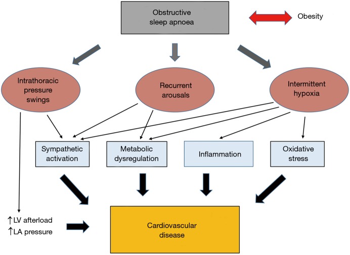
Obstructive sleep apnoea (OSA) is recognized as a major public health burden conveying a significant risk of cardiovascular diseases (CVD) and mortality. Continuous positive airway pressure (CPAP) is the treatment of choice for the majority of patients with OSA but the benefit of CPAP on CVD is uncertain. Thus, a greater understanding of the mechanisms by which OSA leads to CVD might identify novel therapeutic approaches. Intermittent hypoxia (IH), a hallmark feature of OSA, plays a key role in the pathogenesis and experimental studies using animal and cell culture studies suggest that IH mediates CVD through activation of multiple mechanistic pathways such as sympathetic excitation, inflammation, oxidative stress or metabolic dysregulation. Recurrent arousals, intrathoracic pressure swings and concomitant obesity likely play important additive roles in this process. In this review, the available evidence of the pathophysiological mechanisms of CVD in OSA is explored with a specific emphasis on IH, recurrent arousals and intrathoracic pressure swings as the main pathophysiological triggers.
Keywords: Obstructive sleep apnoea (OSA); cardiovascular disease; intermittent hypoxia (IH); sleep fragmentation.
Conflict of interest statement
Conflicts of Interest: The author has no conflicts of interest to declare.
Figures
References
Publication types
LinkOut - more resources
Full Text Sources