An Unusual Case of Giant Cell Tumor of the Distal Tibia
- PMID: 30687657
- PMCID: PMC6343573
- DOI: 10.13107/jocr.2250-0685.1144
An Unusual Case of Giant Cell Tumor of the Distal Tibia
Abstract
Introduction: Giant cell tumors are common in proximal tibia and distal end radius and have a low tendency to recur. They have been treated successfully with excision and cementing or sandwich bone grafting without recurrence. Here, we present a rare case of giant cell tumor (GCT) of the distal tibia treated successfully with no recurrence at the end of 2 years.
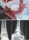
Case report: A 28-year-old female presented with complaints of pain and restricted movement of the right ankle joint since 1 month. There was no history of trauma. On examination, tenderness on the anterior aspect of the right ankle joint with restricted range of motion was found. X-rays revealed a well-defined expansile predominantly lytic lesion in the distal epi-metaphyseal region of the right tibia with minimal periosteal reaction seen along the medial margin. Magnetic resonance imaging revealed an ill-defined expansile lesion involving the epi-metaphyseal end of the lower end of tibia causing cortical breach and having extra-osseous tissue component with the abnormal signal in flexor and extensor group of muscles with the possibility of GCT. Surgery by excision, curettage, and cementation was performed to fill the defect. Histopathology of the tissue showed multinucleated giant cells with a uniform vesicular nucleus and mononuclear cells which were spindle shaped with uniform vesicular nucleus suggestive of GCT.
Conclusion: The patient at 2-year follow-up is doing well, walking without any pain, comfortably and with a full range of motion of the ankle joint with no signs of recurrence.
Keywords: Giant cell tumor distal tibia; curettage for giant cell tumor; giant cell tumor.
Conflict of interest statement
Conflict of Interest: Nil
Figures


References
-
- Turcotte RE. Giant cell tumor of bone. Orthop Clin North Am. 2006;37:35–51. - PubMed
-
- Eckardt JJ, Grogan TJ. Giant cell tumor of bone. Clin Orthop Relat Res. 1986;204:45–58. - PubMed
-
- McGrath PJ. Giant-cell tumour of bone:An analysis of fifty-two cases. J Bone Joint Surg Br. 1972;54:216–29. - PubMed
-
- Bertoni F, Present D, Sudanese A, Baldini N, Bacchini P, Campanacci M, et al. Giant-cell tumor of bone with pulmonary metastases. Six case reports and a review of the literature. Clin Orthop Relat Res. 1988;237:275–85. - PubMed
-
- Siebenrock KA, Unni KK, Rock MG. Giant-cell tumour of bone metastasising to the lungs. A long-term follow-up. J Bone Joint Surg Br. 1998;80:43–7. - PubMed
Publication types
LinkOut - more resources
Full Text Sources
