

Latest advances in intervertebral disc development and progenitor cells
- PMID: 30687811
- PMCID: PMC6338208
- DOI: 10.1002/jsp2.1030
Latest advances in intervertebral disc development and progenitor cells
Abstract
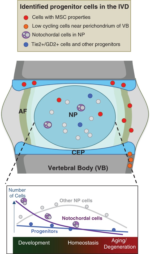
This paper is a concise review aiming to assemble the most relevant topics presented by the authors at ORS-Philadelphia Spine Research Society Fourth International Spine Research Symposium. It centers on the latest advances in disc development, its main structural entities, and the populating cells, with emphasis on the advances in pivotal molecular pathways responsible for forming the intervertebral discs (IVD). The objective of finding and emphasizing pathways and mechanisms that function to control tissue formation is to identify and to explore modifications occurring during normal aging, disease, and tissue repair. Thus, to comprehend that the cellular and molecular basis of tissue degeneration are crucial in the study of the dynamic interplay that includes cell-cell communication, gene regulation, and growth factors required to form a healthy and functional tissue during normal development.
Keywords: development; stem cell; tissue‐specific progenitor cells.
Conflict of interest statement
The authors declare not to have any conflict of interest.
Figures

References
-
- Niehrs C. Regionally specific induction by the Spemann‐Mangold organizer. Nat Rev Genet. 2004;5(6):425‐434. - PubMed
-
- Mangold H. Über die Induktion von Embryonalanlagen durch Implantation artfremder Organisatoren. Roux Arch Entw Mech Org. 1924;100:599‐638.
-
- Oppenheimer JM. Transplantation experiments on developing teleosts (Fundulus and Perca). J Exp Zool. 1936;72:409‐437.
-
- Waddington CH. Developmental mechanics of chick and duck embryos. Nature. 1930;125:924‐925.
-
- Beddington RS. Induction of a second neural axis by the mouse node. Development. 1994;120(3):613‐620. - PubMed
Publication types
Grants and funding
LinkOut - more resources
Full Text Sources
