Case Reports
. 2019 Mar;7(3):e552.
doi: 10.1002/mgg3.552. Epub 2019 Jan 28.

Identification of a pathogenic mutation in ATP2A1 via in silico analysis of exome data for cryptic aberrant splice sites

Affiliations
Case Reports

Identification of a pathogenic mutation in ATP2A1 via in silico analysis of exome data for cryptic aberrant splice sites

Christine C Bruels et al. Mol Genet Genomic Med. 2019 Mar.

Abstract

Background: Pathogenic mutations causing aberrant splicing are often difficult to detect. Standard variant analysis of next-generation sequence (NGS) data focuses on canonical splice sites. Noncanonical splice sites are more difficult to ascertain.

Methods: We developed a bioinformatics pipeline that screens existing NGS data for potentially aberrant novel essential splice sites (PANESS) and performed a pilot study on a family with a myotonic disorder. Further analyses were performed via qRT-PCR, immunoblotting, and immunohistochemistry. RNAi knockdown studies were performed in Drosophila to model the gene deficiency.

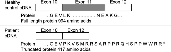
Results: The PANESS pipeline identified a homozygous ATP2A1 variant (NC_000016.9:g.28905928G>A; NM_004320.4:c.1287G>A:p.(Glu429=)) that was predicted to cause the omission of exon 11. Aberrant splicing of ATP2A1 was confirmed via qRT-PCR, and abnormal expression of the protein product sarcoplasmic/endoplasmic reticulum Ca++ ATPase 1 (SERCA1) was demonstrated in quadriceps femoris tissue from the proband. Ubiquitous knockdown of SERCA led to lethality in Drosophila, as did knockdown targeting differentiating or fusing myoblasts.

Conclusions: This study confirms the potential of novel in silico algorithms to detect cryptic mutations in existing NGS data; expands the phenotypic spectrum of ATP2A1 mutations beyond classic Brody myopathy; and suggests that genetic testing of ATP2A1 should be considered in patients with clinical myotonia.

Keywords: ATP2A1; Aberrant RNA splicing; Brody myopathy; cryptic variants.


Conflict of interest statement

The authors declare no conflict of interest.

Figures

Figure 1
Figure 1
The PANESS pipeline accepts input from a VCF or other variant data file. ANNOVAR is used to determine minor allele frequency (MAF) and gene orientation. Low‐frequency variants (MAF ≤ 0.001) are identified and sequence surrounding each variant is extracted. The definition of donor and acceptor essential splice sites on the appropriate strand is used to determine whether each variant creates a new splice site. Lists of candidate PANESS and the affected genes are created
Figure 2
Figure 2
Histological images of quadriceps muscle biopsy on proband 1,406‐1. (a) Hematoxylin and eosin stain shows excessive variation in fiber diameter with both hypertrophic and atrophic fibers and increased centralized nuclei. There is no inflammation and no frank degeneration. There is minimal increased connective tissue. Scale bar, 200 microns. (b) Cytochrome oxidase histochemistry shows a normal stippled pattern, with central pallor suggestive of cores or targets. Scale bar, 100 microns. (c) NADH histochemistry shows a normal stippled pattern, with central pallor suggestive of cores or targets. Scale bar, 100 microns. (d) ATPase stain at pH 9.4 shows mild fiber type grouping of both type 1 and type 2 fibers; the majority of small angulated atrophic fibers are type 2. Scale bar, 200 microns. (e–h) On electron microscopy, the thick and thin filaments, Z lines, M lines, A and I bands are present and generally well‐aligned. Myofiber nuclei, sarcolemmal membranes, and basement membranes are intact. However, there are foci, some quite large, of myofibrillar disorganization and loss of Z‐band structure. Elsewhere, there is streaming of the Z lines and myofibrillar disarray. Nemaline rods are not present, but there is the suggestion that the actin filaments may in some places insert into malformed Z‐band material. There is no increased accumulation of glycogen, abnormal lipid, or other storage material. The mitochondria are normal in number, distribution, and morphology without paracrystalline, osmophilic inclusions, or concentric cristae. There are no vacuolar or degenerative changes in the myofibers
Figure 3
Figure 3
Analysis of family 1406. (a) A family pedigree illustrates the source of consanguinity; the family is from Colombia, and the great‐grandmothers of the proband are sisters. (b) Sanger sequencing of ATP2A1 (NC_000016.9) confirmed a homozygous ATP2A1:c.1287G>A variant (NC_000016.9:g.28905928G>A; NM_004320.4:c.1287G>A:p.(Glu429=)) in 1406‐1 (chromatograms 1 & 2) and a heterozygous ATP2A1:c.1287G>A variant in 1406‐2 and 1406‐3 (chromatograms 3–6). (c) Primers ATP2A1_exon10F1 and ATP2A1_exon12R2 were used to amplify exons 10–12 in cDNA from 1406‐1 (proband) myoblasts (lane 2), along with cDNA (lane 3) and gDNA (lane 4) from healthy control myoblasts. The 1406‐1 amplicon is approximately 100 bp smaller than the control amplicon on a 2% agarose gel. Lanes 1 and 6 show the 2‐log DNA ladder (New England Biolabs), and lane 5 shows a negative control. (d) Sanger sequencing confirmed the omission of exon 11 in cDNA from patient‐derived myoblasts
Figure 4
Figure 4
Diagram represents the predicted effect of skipping exon 11 in ATP2A1 (NC_000016.9) on the SERCA1 amino acid sequence. The variant is the last nucleotide at the 3’ end of exon 11 of ATP2A1 (NC_000016.9:g.28905928G>A; NM_004320.4:c.1287G>A:p.(Glu429=)). It is predicted to affect splicing through the modification of the canonical donor site (splicing signal decreased by 74%), creation of a cryptic acceptor site (splicing signal increased by 66%), and disruption of an exonic splice enhancer site. The predicted omission of exon 11 was confirmed experimentally at the RNA level. The absence of full length and truncated SERCA1 protein expression in the patient is likely due to nonsense‐mediated decay of the mRNA
Figure 5
Figure 5
Analysis of mRNA and protein from proband 1406‐1 and a healthy control. (a) qRT‐PCR of cDNA with probes spanning ATP2A1 (NC_000016.9) exons 9 and 10 show reduced expression of exons 9 and 10 in 1406‐1 compared to control. (b) qRT‐PCR with probes spanning ATP2A1 exons 11 and 12 confirmed undetectable expression of exons 11 and 12 in 1406‐1 compared to control. (c) Representative western blot of SERCA1 protein in control and 1406‐1 quadriceps femoris muscle. The anti‐SERCA1 antibody (Cell Signaling) targeting an epitope surround Leu24 near the N‐terminus of human SERCA1 detected a band of ~100 kDa in control, but no SERCA1 expression in 1406‐1 (left). The SERCA1 expression pattern was confirmed by another anti‐SERCA1 antibody (Developmental Studies Hybridoma Bank) with the same amount of protein loaded (right). (d) Immunofluorescence analysis of laminin (red) and SERCA1 (green) in quadriceps cross‐section from control and 1406‐1. Nuclei were stained with DAPI. Scale bar: 50 μm. Control muscle tissue displays SERCA1 expression most prominently in fast‐twitch fibers, whereas SERCA1 expression is completely absent in muscle tissue obtained from 1406‐1
Figure 6
Figure 6
Adult Drosophila that downregulate SERCA in differentiated muscles display marked defect in locomotor activity. (a) Representative western blot and (b) relative quantification of SERCA protein from control sibling flies (abbreviated genotype CyO; UAS‐ds‐SERCA) and SERCA RNAi flies (abbreviated genotype Mhc‐Gal4; UAS‐ds SERCA). Western blot showing knockdown of SERCA in whole Drosophila lysates. (c, d) Histological analysis of 40 day‐old Mhc‐Gal4>UAS‐ds‐SERCA RNAi flies and CyO; UAS‐ds‐SERCA control siblings. (c) None of the control flies showed the muscle defects (n = 17). Five representative control flies are shown (10× magnification), as well as magnified muscle fibers (inset, 40×). (d) Severe alterations are seen in the large thoracic muscles of SERCA RNAi flies, including the indirect flight muscles and jump muscles (arrows). All SERCA RNAi flies but one display widespread muscle breakdown (n = 15). Other thoracic organs that are readily visible in some samples appear intact (single arrow head, midgut; double arrow head, ventral nerve cord). Five representative flies are shown (10× magnification), as well as magnified muscle fibers (inset, 40×). (e–h) Adult SERCA RNAi and control sibling flies were collected and functionally characterized. (e–g) In each picture, the left vial contains the control flies, and the right vial contains the SERCA RNAi flies. (e) The flies were prompted by tapping both vials simultaneously once on a flat surface. (f–g) The climbing ability of the flies was video recorded. Representative still photographs are shown. Most control siblings reach the top in <10 s, while SERCA RNAi flies move markedly more slowly. The corresponding video can be accessed in the Supporting information Video S1. (h) Negative geotaxis assay. Control siblings (n = 10–14) and SERCA RNAi flies (n = 9–13) were assessed for their ability to cross a 3‐cm‐threshold line in 12 s. The assay was performed in triplicate and repeated three times at 1‐min intervals (9 times total). Few SERCA RNAi flies were able to reach the line in the given time compared to control siblings

Similar articles

Cited by

References

    1. Aarskog, N. K. , & Vedeler, C. A. (2000). Real‐time quantitative polymerase chain reaction. A new method that detects both the peripheral myelin protein 22 duplication in Charcot‐Marie‐Tooth type 1A disease and the peripheral myelin protein 22 deletion in hereditary neuropathy with liability to pressure palsies. Human Genetics, 107(5), 494–498. 10.1007/s004390000399 - DOI - PubMed
    1. Abraham, D. M. , & Wolf, M. J. (2013). Disruption of sarcoendoplasmic reticulum calcium ATPase function in Drosophila leads to cardiac dysfunction. PLoS One, 8(10), e77785 10.1371/journal.pone.0077785. - DOI - PMC - PubMed
    1. Alexander, M. S. , Casar, J. C. , Motohashi, N. , Myers, J. A. , Eisenberg, I. , Gonzalez, R. T. , … Kunkel, L. M. (2011). Regulation of DMD pathology by an ankyrin‐encoded miRNA. Skeletal Muscle, 1, 27 10.1186/2044-5040-1-27. - DOI - PMC - PubMed
    1. Ankala, A. , da Silva, C. , Gualandi, F. , Ferlini, A. , Bean, L. J. , Collins, C. , … Hegde, M. R. (2015). A comprehensive genomic approach for neuromuscular diseases gives a high diagnostic yield. Annals of Neurology, 77(2), 206–214. 10.1002/ana.24303. - DOI - PubMed
    1. Bonne, G. , Rivier, F. , & Hamroun, D. (2017). The 2018 version of the gene table of monogenic neuromuscular disorders (nuclear genome). Neuromuscular Disorders, 27(12), 1152–1183. 10.1016/j.nmd.2017.10.005. - DOI - PubMed

Publication types

Substances

LinkOut - more resources