

Combined cellular and soluble mediator analysis for improved diagnosis of vitreoretinal lymphoma
- PMID: 30688042
- PMCID: PMC6796208
- DOI: 10.1111/aos.14036
Combined cellular and soluble mediator analysis for improved diagnosis of vitreoretinal lymphoma
Abstract
Purpose: Primary vitreoretinal lymphoma [(P)VRL]) is a rare malignancy of the eye localized in the retina, vitreous or choroid. Here, we aim to determine the value of the combination of innovative diagnostic methods for accurate differentiation between (P)VRL and non-(P)VRL in patients with suspect uveitis or vitritis.
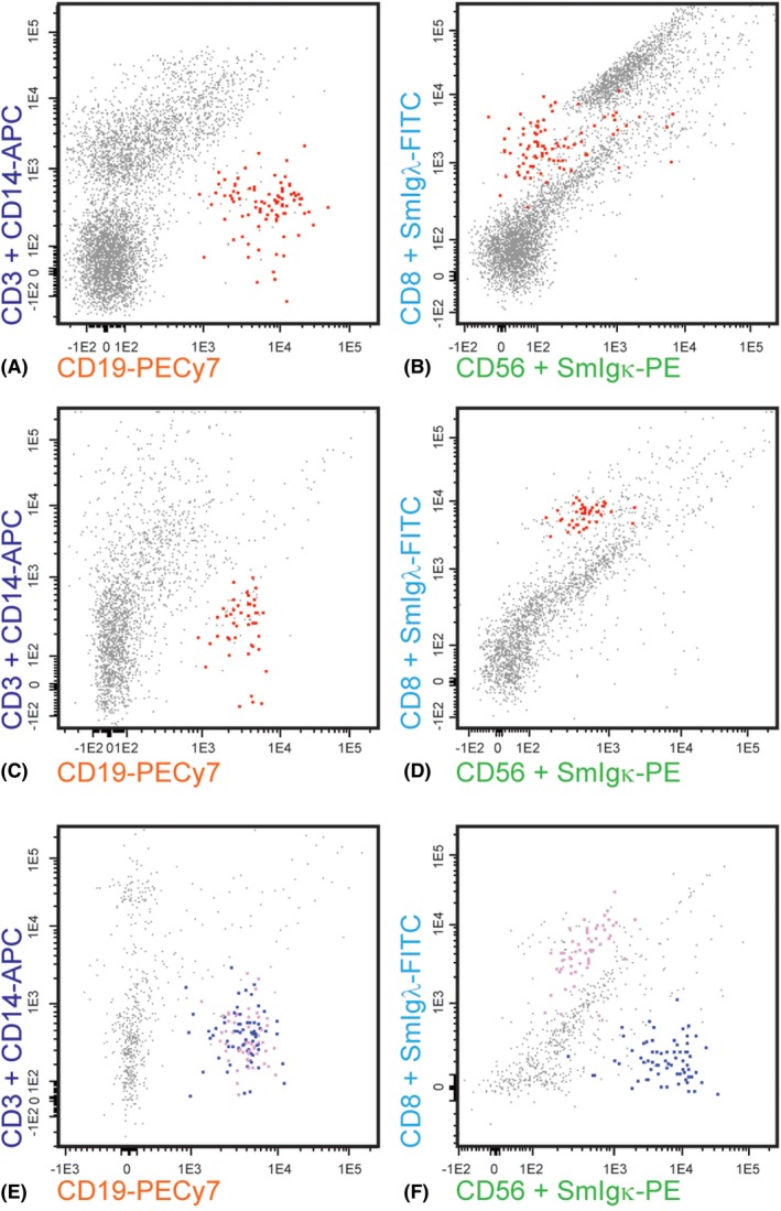
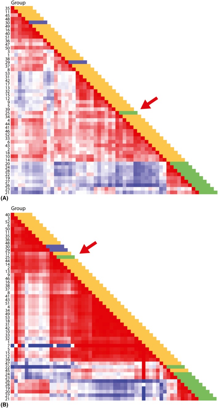
Methods: Multicolour flow cytometric immunophenotyping of cells in the vitreous samples was performed using the EuroFlow small sample tube. Additionally, cytokines/chemokines and growth factors were measured in the vitreous specimens using a multiplex immunoassay. Data were evaluated in predefined clinical subgroups using omniviz unsupervised Pearson's correlation visualization and unsupervised heatmap analysis.
Results: A total of 53 patients were prospectively included in the period 2012-2015. In the (P)VRL subgroup (n = 10), nine cases showed aberrant surface membrane immunoglobulin (SmIg) light chain expression. In the non-(P)VRL group (n = 43) clearly skewed SmIg light chain expression was observed in two multiple sclerosis-related uveitis cases, but not in other uveitis types. Soluble mediator measurement revealed high interleukin (IL)-10/IL-6 ratios, and high IL-1RA levels in 9/10 (P)VRL cases, but not in any non-(P)VRL case. Further correlation and heatmap analysis revealed a minimal signature of cellular parameters (CD19+ B cells, aberrant SmIg light chain expression) and cytokine parameters (IL-10/IL-6 ratio >1, high IL-10, high IL-1 RA, high monocyte chemotactic protein-1, high macrophage inflammatory protein-1β) to reliably distinguish (P)VRL from non-(P)VRL.
Conclusion: Here, we show the power of a combined cellular and proteomics strategy for detecting (P)VRL in vitreous specimens, especially in cases with minor cellular (P)VRL infiltrates.
Keywords: diagnostics; immunology; multiparameter flow cytometry; soluble mediators; uveitis; vitreoretinal lymphoma.
© 2019 Acta Ophthalmologica Scandinavica Foundation. Published by John Wiley & Sons Ltd.
Figures
References
-
- Bastiaans J, van Meurs JC, Mulder VC et al. (2014): The role of thrombin in proliferative vitreoretinopathy. Invest Ophthalmol Vis Sci 55: 4659–4666. - PubMed
-
- Bonzheim I, Giese S, Deuter C et al. (2015): High frequency of MYD88 mutations in vitreoretinal B‐cell lymphoma: a valuable tool to improve diagnostic yield of vitreous aspirates. Blood 126: 76–79. - PubMed
-
- Buggage RR, Whitcup SM, Nussenblatt RB & Chan CC (1999): Using interleukin 10 to interleukin 6 ratio to distinguish primary intraocular lymphoma and uveitis. Invest Ophthalmol Vis Sci 40: 2462–2463. - PubMed
MeSH terms
Substances
Grants and funding
LinkOut - more resources
Full Text Sources
Medical
Research Materials
