. 2019 May;14(5):775-782.
doi: 10.4103/1673-5374.249225.

Early constraint-induced movement therapy affects behavior and neuronal plasticity in ischemia-injured rat brains

Affiliations

Early constraint-induced movement therapy affects behavior and neuronal plasticity in ischemia-injured rat brains

Xi-Hua Liu et al. Neural Regen Res. 2019 May.

Abstract

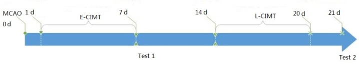
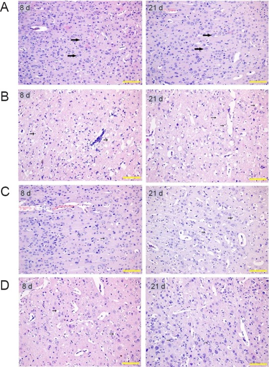
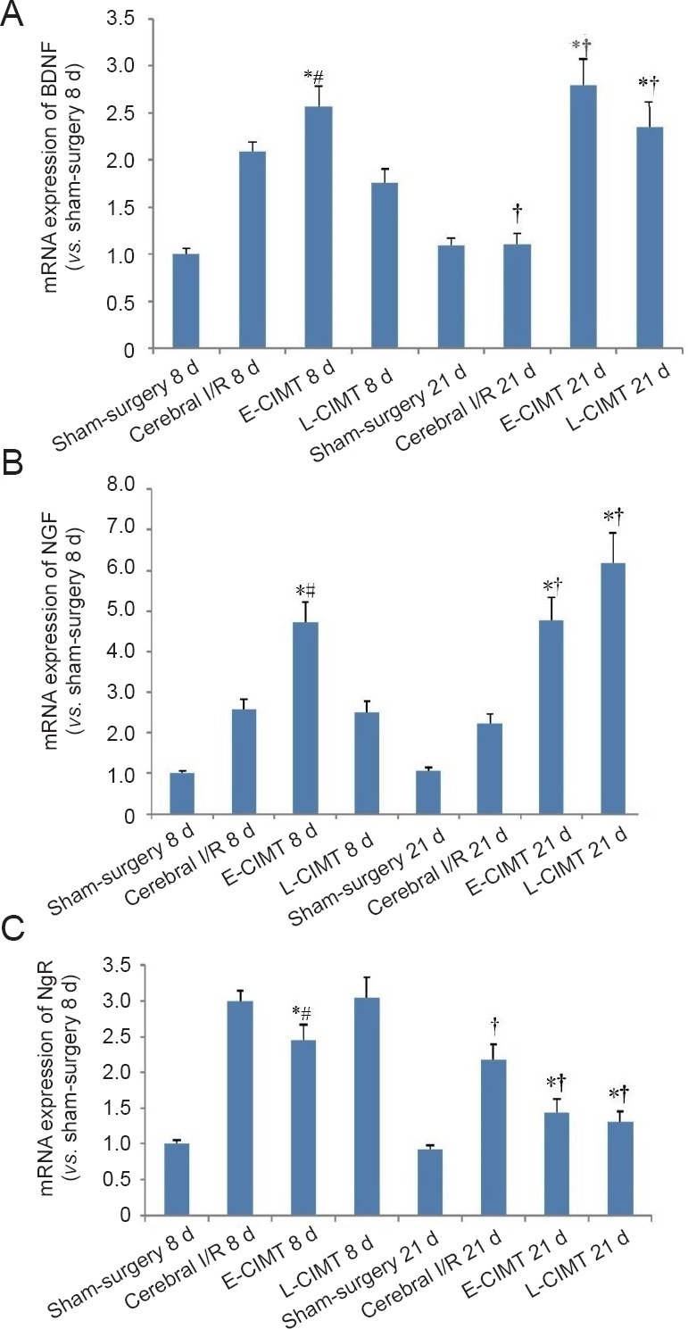
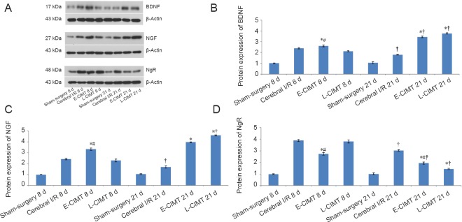
Constraint-induced movement therapy is an effective rehabilitative training technique used to improve the restoration of impaired upper extremity movement after stroke. However, whether constraint-induced movement therapy is more effective than conventional rehabilitation in acute or sub-acute stroke remains controversial. The aim of the present study was to identify the optimal time to start constraint-induced movement therapy after ischemic stroke and to explore the mechanisms by which constraint-induced movement therapy leads to post-stroke recovery. Sixty-four adult male Sprague-Dawley rats were randomly divided into four groups: sham-surgery group, cerebral ischemia/reperfusion group, early constraint-induced movement therapy group, and late constraint-induced movement therapy group. Rat models of left middle cerebral artery occlusion were established according to the Zea Longa line embolism method. Constraint-induced movement therapy was conducted starting on day 1 or day 14 in the early constraint-induced movement therapy and late constraint-induced movement therapy groups, respectively. To explore the effect of each intervention time on neuromotor function, behavioral function was assessed using a balance beam walking test before surgery and at 8 and 21 days after surgery. The expression levels of brain-derived neurotrophic factor, nerve growth factor and Nogo receptor were evaluated using real time-polymerase chain reaction and western blot assay to assess the effect of each intervention time. The results showed that the behavioral score was significantly lower in the early constraint-induced movement therapy group than in the cerebral ischemia/reperfusion and late constraint-induced movement therapy groups at 8 days. At 21 days, the scores had significantly decreased in the early constraint-induced movement therapy and late constraint-induced movement therapy groups. At 8 days, only mild pyknosis appeared in neurons of the ischemic penumbra in the early constraint-induced movement therapy group, which was distinctly better than in the cerebral ischemia/reperfusion group. At 21 days, only a few vacuolated cells were observed and no obvious inflammatory cells were visible in late constraint-induced movement therapy group, which was much better than at 8 days. The mRNA and protein expression levels of brain-derived neurotrophic factor and nerve growth factor were significantly higher, but expression levels of Nogo receptor were significantly lower in the early constraint-induced movement therapy group compared with the cerebral ischemia/reperfusion and late constraint-induced movement therapy groups at 8 days. The changes in expression levels at 21 days were larger but similar in both the early constraint-induced movement therapy and late constraint-induced movement therapy groups. Besides, the protein nerve growth factor level was higher in the late constraint-induced movement therapy group than in the early constraint-induced movement therapy group at 21 days. These results suggest that both early (1 day) and late (14 days) constraint-induced movement therapy induces molecular plasticity and facilitates functional recovery after ischemic stroke, as illustrated by the histology. The mechanism may be associated with downregulation of Nogo receptor expression and upregulation of brain-derived neurotrophic factor and nerve growth factor expression.

Keywords: constraint-induced movement therapy; functional recovery; ischemic stroke; nerve growth factors; nerve regeneration; neural regeneration; neuronal plasticity; rats; real time-polymerase chain reaction; rehabilitation; western blot assay.


Conflict of interest statement

None

Figures

Figure 1
Figure 1
Protocol of the present study. Cerebral infarction model rats were made according to the Zea Longa line embolism method, followed by 7 days CIMT from day 1 or day 14. Behavioral assessment by neurological score and balance beam walking test was carried out after CIMT (test 1 at day 8, and test 2 at day 21). Hematoxylin-eosin staining, real-time polymerase chain reaction and western blot assay for Nogo-receptor, brain-derived neurotrophic factor and nerve growth factor were also performed after early and late CIMT. E-CIMT: Early CIMT, forced-limb use of the affected forelimb for 7 days, starting on day 1 following ischemic stroke; L-CIMT: late CIMT, forced-limb use of the affected forelimb for 7 days, starting on day 14 following ischemic stroke; MCAO: middle cerebral artery occlusion; CIMT: constraint-induced movement therapy; d: day(s).
Figure 2
Figure 2
Effect of CIMT on histological changes in rats with ischemic brain injury (hematoxylin-eosin staining). (A) Histological changes of sham-surgery group at 8 and 21 days: No obvious lesions at 8 and 21 days. The nucleus was rounded; the nucleolus was clear; the intercellular space was normal. (B) Histological changes of cerebral ischemia/reperfusion group at 8 and 21 days: At 8 days, obvious pyknosis, cytoplasmic vacuolization and vacuolar degeneration were observed. Neuronal number decreased and intercellular space increased. At 21 days, the neuropile became very rarefacted and glial cells increased in addition to the pathological changes at 8 days. (C) Histological changes of E-CIMT group at 8 and 21 days: At 8 days, the mild pyknosis of neurons seen in the ischemic penumbra in E-CIMT group was significantly less than in the cerebral ischemia/reperfusion group. By 21 days, the severity of the lesions showed improvement compared with that at 8 days, but much less than in the ischemia/reperfusion only group. (D) Histological changes of L-CIMT group at 8 and 21 days: At 8 days, the detrimental histological changes from both the L-CIMT and cerebral ischemia/reperfusion groups were more severe than in the E-CIMT group. At 21 days, the severity in the L-CIMT group was slightly less than in the E-CIMT group. Obvious pyknosis and few vacuolated cells were also observed. Original magnification: 200×. Scale bars: 20 μm. E-CIMT: Early CIMT, forced-limb use of the affected forelimb for 7 days, starting on day 1 following ischemic stroke; L-CIMT: late CIMT, forced-limb use of the affected forelimb for 7 days, starting on day 14 following ischemic stroke; CIMT: constraint-induced movement therapy; d: days.
Figure 3
Figure 3
CIMT effect on mRNA expression levels of BDNF, NGF, and NgR in rats with ischemic brain injury. (A) At 8 days, mRNA expression levels of BDNF were higher in the E-CIMT group than in the cerebral I/R and L-CIMT groups. The expression levels of BDNF had significantly increased at 21 days compared with those at 8 days in the L-CIMT group using 2-ΔΔCT method. The expression levels of BDNF were higher in the E-CIMT and L-CIMT groups than in the cerebral I/R group. (B) Results of mRNA expression of NGF were similar to those for BDNF. (C) At 8 days, the E-CIMT group exhibited remarkably lower mRNA expression of NgR compared with the cerebral I/R and L-CIMT groups. The mRNA expression levels of NgR had significantly decreased at 21 days compared with those at 8 days in the cerebral I/R, E-CIMT, and L-CIMT groups. The expression levels in E-CIMT and L-CIMT groups were lower than that in the cerebral I/R group. Data are expressed as the mean ± SEM (n = 8; one-way analysis of variance followed by Student-Newman-Keuls post hoc test). *P < 0.05, vs. cerebral I/R group; #P < 0.05, vs. L-CIMT group; †P < 0.05, vs. 8 days. E-CIMT: Early CIMT, forced-limb use of the affected forelimb for 7 days, starting on day 1 following ischemic stroke; L-CIMT: late CIMT, forced-limb use of the affected forelimb for 7 days, starting on day 14 following ischemic stroke. BDNF: Brain-derived neurotrophic factor; NGF: nerve growth factor; NgR: Nogo-receptor; CIMT: constraint-induced movement therapy; I/R: ischemia/reperfusion; d: days.
Figure 4
Figure 4
CIMT effect on protein expression levels of BDNF, NGF and NgR in rats with ischemic brain injury. (A) Protein expression levels of BDNF, NGF and NgR in different groups with β-actin as the internal control. (B) Western blot assay for BDNF protein expression at 8 and 21 days: BDNF expression was significantly higher in the E-CIMT group than in the cerebral I/R and L-CIMT groups at day 8. The relative expression of BDNF was increased at 21 days compared with that at 8 days in the E-CIMT and L-CIMT groups. BDNF levels in the E-CIMT and L-CIMT groups were significantly higher than in the cerebral I/R and sham-surgery groups (P < 0.05). (C) Protein expression of NGF at 8 and 21 days: The changes in protein expression of NGF were similar to those of BDNF. (D) Protein expression of NgR at 8 and 21 days: At 8 days, the NgR level in the E-CIMT group was significantly lower than in the cerebral I/R and L-CIMT groups. At day 21 NgR expression was significantly reduced compared with that at 8 days in the cerebral I/R, E-CIMT and L-CIMT groups. Expressions of NgR in the E-CIMT and L-CIMT groups were lower than in the cerebral I/R group. Data are expressed as the mean ± SEM (n = 8; one-way analysis of variance followed by Student-Newman-Keuls post hoc test). *P < 0.05, vs. cerebral I/R group; #P < 0.05, vs. L-CIMT group; †P < 0.05, vs. 8 days. E-CIMT: Early CIMT, forced-limb use of the affected forelimb for 7 days, starting on day 1 following ischemic stroke; L-CIMT: late CIMT, forced-limb use of the affected forelimb for 7 days, starting on day 14 following ischemic stroke. BDNF: Brain-derived neurotrophic factor; NGF: nerve growth factor; NgR: Nogo-receptor; CIMT: constraint-induced movement therapy; I/R: ischemia/reperfusion; d: days.

Similar articles

Cited by

References

    1. Azab M, Al-Jarrah M, Nazzal M, Maayah M, Sammour MA, Jamous M. Effectiveness of constraint-induced movement therapy (CIMT) as home-based therapy on Barthel Index in patients with chronic stroke. Top Stroke Rehabil. 2009;16:207–211. - PubMed
    1. Bachman J. Reverse-transcription PCR (RT-PCR) Methods Enzymol. 2013;530:67–74. - PubMed
    1. Berretta A, Tzeng YC, Clarkson AN. Post-stroke recovery: the role of activity-dependent release of brain-derived neurotrophic factor. Expert Rev Neurother. 2014;14:1335–1344. - PubMed
    1. Cheatwood JL, Emerick AJ, Schwab ME, Kartje GL. Nogo-A expression after focal ischemic stroke in the adult rat. Stroke. 2008;39:2091–2098. - PMC - PubMed
    1. Cheng S, Ma M, Ma Y, Wang Z, Xu G, Liu X. Combination therapy with intranasal NGF and electroacupuncture enhanced cell proliferation and survival in rats after stroke. Neurol Res. 2009;31:753–758. - PubMed