. 2019 Jan 11:(143):10.3791/58461.
doi: 10.3791/58461.

Use of Electron Paramagnetic Resonance in Biological Samples at Ambient Temperature and 77 K

Affiliations

Use of Electron Paramagnetic Resonance in Biological Samples at Ambient Temperature and 77 K

Hanan B Elajaili et al. J Vis Exp. .

Abstract

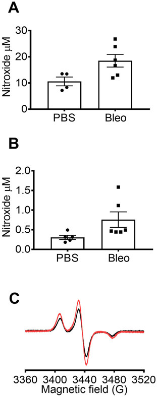
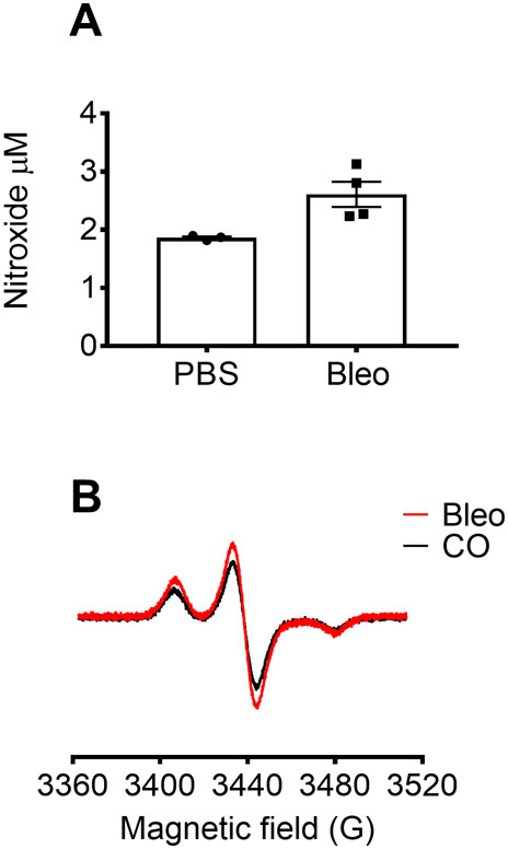
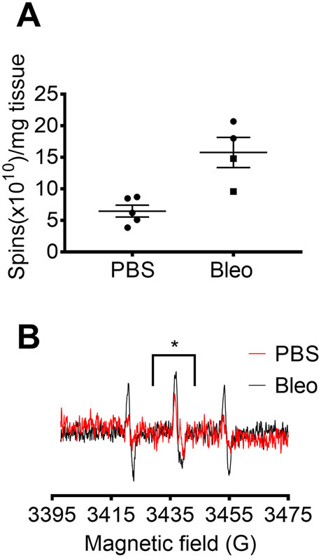
The accurate and specific detection of reactive oxygen species (ROS) in different cellular and tissue compartments is essential to the study of redox-regulated signaling in biological settings. Electron paramagnetic resonance spectroscopy (EPR) is the only direct method to assess free radicals unambiguously. Its advantage is that it detects physiologic levels of specific species with a high specificity, but it does require specialized technology, careful sample preparation, and appropriate controls to ensure accurate interpretation of the data. Cyclic hydroxylamine spin probes react selectively with superoxide or other radicals to generate a nitroxide signal that can be quantified by EPR spectroscopy. Cell-permeable spin probes and spin probes designed to accumulate rapidly in the mitochondria allow for the determination of superoxide concentration in different cellular compartments. In cultured cells, the use of cell permeable 1-hydroxy-3-methoxycarbonyl-2,2,5,5-tetramethylpyrrolidine (CMH) along with and without cell-impermeable superoxide dismutase (SOD) pretreatment, or use of cell-permeable PEG-SOD, allows for the differentiation of extracellular from cytosolic superoxide. The mitochondrial 1-hydroxy-4-[2-triphenylphosphonio)-acetamido]-2,2,6,6-tetramethyl-piperidine,1-hydroxy-2,2,6,6-tetramethyl-4-[2-(triphenylphosphonio)acetamido] piperidinium dichloride (mito-TEMPO-H) allows for measurement of mitochondrial ROS (predominantly superoxide). Spin probes and EPR spectroscopy can also be applied to in vivo models. Superoxide can be detected in extracellular fluids such as blood and alveolar fluid, as well as tissues such as lung tissue. Several methods are presented to process and store tissue for EPR measurements and deliver intravenous 1-hydroxy-3-carboxy-2,2,5,5-tetramethylpyrrolidine (CPH) spin probe in vivo. While measurements can be performed at room temperature, samples obtained from in vitro and in vivo models can also be stored at -80 °C and analyzed by EPR at 77 K. The samples can be stored in specialized tubing stable at -80 °C and run at 77 K to enable a practical, efficient, and reproducible method that facilitates storing and transferring samples.


Figures

Figure 1:
Figure 1:. Detection of superoxide in different cell compartments.
(A) EPR spectra generated by 0.25 mM CMH in 0.5 mM hypoxanthine/xanthine oxidase (8 mU/mL) with and without SOD (30 U/mL). (B) RAW 264.7 cells (1 x 10⁶ cells/well) were stimulated with 10 μM PMA in the presence of CMH for 50 min at 37 °C and nitroxide concentration (μM) detected in cell suspension (cells + buffer) and buffer collected from treated cells. (C) RAW 264.7 cells were stimulated with PMA vs. vehicle control (Con). One set of cells were pretreated for 10 min with 30 U/mL cell-impermeable SOD (PMA + SOD). Each color represents data from different experimental days and each point represents cells from an individual well. The nitroxide signal in a time-matched blank with CMH in KHB was subtracted from each signal to obtain final values. (D) Calculation of total and extracellular superoxide in PMA stimulated cells; T = total superoxide, EC = extracellular superoxide (SOD inhibitable signal). (E) To evaluate the intracellular superoxide signal (IC), the signal in buffer after PMA + SOD was compared to PMA-treated cells after the removal of buffer. To confirm, wells were pretreated with 60 U/mL cell-permeable PEG-SOD for 1.5 hours to determine the intracellular SOD inhibitable. The time-matched CMH blank is shown, and data reflect absolute nitroxide signal. Data expressed as mean ± SEM.
Figure 2:
Figure 2:. Detection of mitochondrial superoxide in RAW cells stimulated with antimycin A.
(A) Representative spectra of the mitochondrial-specific EPR spin probe, 0.25 mM mito-TEMPO-H in RAW 264.7 cells without (Con) or with 25 μM antimycin A (AA) for 50 min at 37 °C. (B) CM. concentration (μM) in cells treated with AA compared to control. The nitroxide signal in a time-matched mito-TEMPO-H blank was subtracted from total signal to obtain final values. Data expressed as mean ± SEM.
Figure 3:
Figure 3:. Detection of superoxide in RAW 264.7 cells at 77K.
(A) RAW 264.7 cells stimulated with 10 μM PMA and EPR spin probe, CMH 0.25 mM (50 min at 37 °C) with (black) or without (red) pretreatment with 30 U/mL SOD. 100 μL of supernatant was loaded in a 1-inch in length piece of PTFE tubing, then flash frozen in liquid nitrogen. The stoppers were removed, and frozen PTFE tubing was placed in the finger dewar for data acquisition at 77 K. (B) A photo of PTFE tubing and stoppers.
Figure 4:
Figure 4:. EPR measurements in blood and BALF from control and bleomycin-treated mice.
Mice were treated with a single dose of intratracheal bleomycin (IT Bleo) (100 μL at 1 U/mL) or PBS vehicle. At 7 days, mice were anesthetized and euthanized. Blood was collected via right ventricular puncture into a syringe coated with 1000 USP/mL heparin containing 100 μM DTPA. Bronchoalveolar lavage fluid (BALF) was collected by lavaging the lungs with 1 mL of 100 μM DTPA in PBS. Blood and BALF were incubated for 10 or 50 min, respectively, with 0.2 mM CMH at 37 °C. 150 μL of blood or BALF was loaded in PTFE tubing flash frozen in liquid nitrogen and EPR data collected at 77 K using a finger dewar. Data show nitroxide concentrations in (A) blood and (B) BALF from PBS- and Bleo-treated mice (n = 4-6). Data expressed as mean ± SEM. (C) Representative spectra of nitroxide in blood from PBS- and Bleo-treated mice.
Figure 5:
Figure 5:. EPR measurements in flash frozen lung tissue.
Mice were treated with a single dose of intratracheal bleomycin (IT bleo) (100 μL at 1 U/mL) or PBS vehicle. At 7 days, the lungs were flushed with cold PBS to remove blood and flash frozen in liquid nitrogen. 5-15 mg of flash-frozen lung tissue was incubated with 0.2 mM CMH in KHB containing 100 μM in 200 μL of total volume for 1 h at 37° C. Supernatant was collected and placed in PTFE tubing and run at 77 K in the finger dewar. (A) Nitroxide concentration (μM of nitroxide normalized to 1 mg of tissue). Data represent the average of 2-3 measurements for each lung. Data expressed as mean ± SEM. (B) Representative spectra of nitroxide in lung tissue from PBS- and Bleo-treated mice.
Figure 6:
Figure 6:. EPR measurements in lung tissue preserved in sucrose buffer.
Mice were treated with a single dose of intratracheal bleomycin (100 μL at 1 U/mL). At 7 days post-treatment, the lungs were flushed with cold PBS to remove blood, and fresh lung tissue was homogenized in Tris-EDTA buffer containing 0.25 mM sucrose at a 1:6 lung weight/buffer volume (mg/μL) ratio. 50 μL of lung homogenate was preincubated with KHB with or without the following inhibitors for 20 min at 37 °C: SOD (100 U/mL), deferoxamine (DFO; 800 μM), and diphenyliodonium chloride (DIP; 100 μM) followed by incubation with 0.2 mM CMH in KHB containing 100 μM DTPA for 20 min at 37 °C. Data was obtained at RT using EPR capillary tubes. (A) Nitroxide concentration in lungs from PBS- and Bleo-treated mice. (B) Nitroxide concentration in Bleo lungs in the absence or the presence of inhibitors (n=3). Data expressed as mean ± SEM.
Figure 7:
Figure 7:. EPR measurements in lung tissue from mice injected with CPH spin probe.
100 μL of CPH was administered via retroorbital injection for a final concentration of 20 mg of CPH per kg of body weight. After 1 h of circulation, mice were euthanized, lungs were flushed with 10 mL of cold PBS via the right ventricle, and lung tissue was flash frozen. 20 to 30 mg of lung tissue was placed in tissue cell and EPR measurements performed at RT. (A) Data expressed as spins/mg. (B) Representative spectra of nitroxide signal in PBS and Bleo lung tissues (* indicates the overlap with ascorbic acid radical). Data expressed as mean ± SEM.

References

    1. Kalyanaraman B, et al. Measuring reactive oxygen and nitrogen species with fluorescent probes: challenges and limitations. Free Radical Biology and Medicine. 52 (1), 1–6 (2012). - PMC - PubMed
    1. Bobko AA, et al. In vivo monitoring of pH, redox status, and glutathione using L-band EPR for assessment of therapeutic effectiveness in solid tumors. Magnetic Resonance in Medicine. 67 (6), 1827–1836 (2012). - PMC - PubMed
    1. Elajaili HB, et al. Electron spin relaxation times and rapid scan EPR imaging of pH-sensitive amino-substituted trityl radicals. Magnetic Resonance in Chemistry. 53 (4), 280–284 (2015). - PMC - PubMed
    1. Elajaili H, et al. Imaging disulfide dinitroxides at 250 MHz to monitor thiol redox status. Journal of Magnetic Resonance. 260, 77–82 (2015). - PMC - PubMed
    1. Halpern HJ, et al. Oxymetry Deep in Tissues with Low-Frequency Electron-Paramagnetic-Resonance. Proceedings of the National Academy of Sciences of the United States of America. 91 (26), 13047–13051 (1994). - PMC - PubMed

Publication types