

Depth of invasion in patients with early stage oral cancer staged by sentinel node biopsy
- PMID: 30688384
- PMCID: PMC6618049
- DOI: 10.1002/hed.25665
Depth of invasion in patients with early stage oral cancer staged by sentinel node biopsy
Abstract
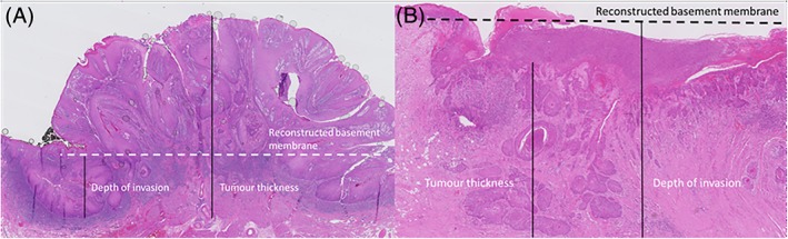
Background: To investigate if depth of invasion (DOI) can predict occult nodal disease in patients with cT1-2N0 (7th TNM) oral squamous cell carcinoma (OSCC) staged by sentinel lymph node biopsy (SLNB).
Methods: In 199 OSCC patients, DOI measurements and SLNB were performed.
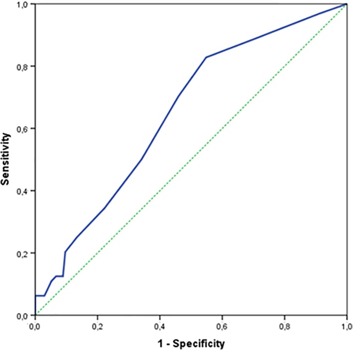
Results: Metastases were found in 64 of 199 patients (32%). Of these 64 patients, the mean DOI was 6.6 mm compared to 4.7 mm in patients without metastases (P = .003). The ROC-curve showed an area under the curve of 0.65 with a most optimal cutoff point of 3.4 mm DOI (sensitivity 83% and specificity 47%). Regional metastases were found in 15% of patients with DOI ≤ 3.4 mm.
Conclusion: DOI seems to be a poor predictor for regional metastasis in patients with cT1-2N0 OSCC. Therefore, staging of the neck using SLNB in patients with early stage oral cancer should also be performed in tumors with limited DOI and probably in T3 (8th TNM) OSCC ≤4 cm diameter.
Keywords: Oral cancer; depth of invasion; lymph node metastases; sentinel lymph node biopsy.
© 2019 The Authors. Head & Neck published by Wiley Periodicals, Inc.
Conflict of interest statement
The authors declare no conflict of interest.
Figures


Similar articles
-
Preoperative assessment of CD44-mediated depth of invasion as predictor of occult metastases in early oral squamous cell carcinoma.Head Neck. 2019 Apr;41(4):950-958. doi: 10.1002/hed.25532. Epub 2018 Dec 18. Head Neck. 2019. PMID: 30561155
-
Sentinel lymph node biopsy for early oral cancer - accuracy and considerations in patient selection.Br J Oral Maxillofac Surg. 2022 Jul;60(6):830-836. doi: 10.1016/j.bjoms.2021.12.058. Epub 2022 Jan 12. Br J Oral Maxillofac Surg. 2022. PMID: 35331563
-
Cytokeratin 19 expression in early oral squamous cell carcinoma and their metastasis: Inadequate biomarker for one-step nucleic acid amplification implementation in sentinel lymph node biopsy procedure.Head Neck. 2017 Sep;39(9):1864-1868. doi: 10.1002/hed.24847. Epub 2017 Jun 23. Head Neck. 2017. PMID: 28644540
-
What is the role of sentinel lymph node biopsy in the management of oral cancer in 2020?Eur Arch Otorhinolaryngol. 2021 Sep;278(9):3181-3191. doi: 10.1007/s00405-020-06538-y. Epub 2020 Dec 28. Eur Arch Otorhinolaryngol. 2021. PMID: 33369691 Free PMC article. Review.
-
Management of the clinically N0 neck in early-stage oral squamous cell carcinoma (OSCC). An EACMFS position paper.J Craniomaxillofac Surg. 2020 Aug;48(8):711-718. doi: 10.1016/j.jcms.2020.06.004. Epub 2020 Jul 2. J Craniomaxillofac Surg. 2020. PMID: 32718880 Review.
Cited by
-
Research on neck dissection for oral squamous-cell carcinoma: a bibliometric analysis.Int J Oral Sci. 2021 Apr 1;13(1):13. doi: 10.1038/s41368-021-00117-5. Int J Oral Sci. 2021. PMID: 33795644 Free PMC article.
-
Addition of tumour infiltration depth and extranodal extension improves the prognostic value of the pathological TNM classification for early-stage oral squamous cell carcinoma.Histopathology. 2019 Sep;75(3):329-337. doi: 10.1111/his.13886. Epub 2019 Jul 29. Histopathology. 2019. PMID: 31021008 Free PMC article.
-
Macrophage CCL22 expression promotes lymphangiogenesis in patients with tongue squamous cell carcinoma via IL-4/STAT6 in the tumor microenvironment.Oncol Lett. 2021 May;21(5):383. doi: 10.3892/ol.2021.12644. Epub 2021 Mar 16. Oncol Lett. 2021. PMID: 33777206 Free PMC article.
-
Comparison of Different Staging Systems Applied to a Cohort of Patients With Oral Tongue and Floor of the Mouth Cancer.Front Oral Health. 2021 Sep 23;2:737329. doi: 10.3389/froh.2021.737329. eCollection 2021. Front Oral Health. 2021. PMID: 35048052 Free PMC article.
-
Accuracy of magnetic resonance imaging in the assessment of depth of invasion in tongue carcinoma: A systematic review and meta-analysis.Natl J Maxillofac Surg. 2023 Sep-Dec;14(3):341-353. doi: 10.4103/njms.njms_174_22. Epub 2023 Nov 10. Natl J Maxillofac Surg. 2023. PMID: 38273911 Free PMC article. Review.
References
-
- Kalnins IK, Leonard AG, Sako K, Razack MS, Shedd DP. Correlation between prognosis and degree of lymph node involvement in carcinoma of the oral cavity. Am J Surg. 1977;134:450‐454. - PubMed
-
- Woolgar JA, Rogers S, West CR, Errington RD, Brown JS, Vaughan ED. Survival and patterns of recurrence in 200 oral cancer patients treated by radical surgery and neck dissection. Oral Oncol. 1999;35:257‐265. - PubMed
-
- Gourin CG, Conger BT, Porubsky ES, Sheils WC, Bilodeau PA, Coleman TA. The effect of occult nodal metastases on survival and regional control in patients with head and neck squamous cell carcinoma. Laryngoscope. 2008;118:1191‐1194. - PubMed
-
- Ganly I, Patel S, Shah J. Early stage squamous cell cancer of the oral tongue‐‐clinicopathologic features affecting outcome. Cancer. 2012;118(1):101‐111. - PubMed
MeSH terms
LinkOut - more resources
Full Text Sources
Medical
