Pilot study of rapid MR pancreas screening for patients with BRCA mutation
- PMID: 30689033
- PMCID: PMC6609466
- DOI: 10.1007/s00330-018-5975-0
Pilot study of rapid MR pancreas screening for patients with BRCA mutation
Abstract
Purpose: To develop and optimize a rapid magnetic resonance imaging (MRI) screening protocol for pancreatic cancer to be performed in conjunction with breast MRI screening in breast cancer susceptibility gene (BRCA)-positive individuals.
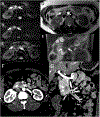
Methods: An IRB-approved prospective study was conducted. The rapid screening pancreatic MR protocol was designed to be less than 10 min to be performed after a standard breast MRI protocol. Protocol consisted of coronal NT T2 SSFSE, axial NT T2 SSFSE and axial NT rFOV FOCUS DWI, and axial T1. Images were acquired with the patient in the same prone position of breast MRI using the built-in body coil. Image quality was qualitatively assessed by two radiologists with 12 and 13 years of MRI experience, respectively. The imaging protocol was modified until an endpoint of five consecutive patients with high-quality diagnostic images were achieved. Signal-to-noise ratio and contrast-to-noise ratio were assessed.
Results: The rapid pancreas MR protocol was successfully completed in all patients. Diagnostic image quality was achieved for all patients. Excellent image quality was achieved for low b values; however, image quality at higher b values was more variable. In one patient, a pancreatic neuroendocrine tumor was found and the patient was treated surgically. In four patients, small pancreatic cystic lesions were detected. In one subject, a hepatic mass was identified and confirmed as adenoma by liver MRI.
Conclusion: Rapid MR protocol for pancreatic cancer screening is feasible and has the potential to play a role in screening BRCA patients undergoing breast MRI.
Key point: • Develop and optimize a rapid magnetic resonance imaging (MRI) screening protocol for pancreatic cancer to be performed in conjunction with breast MRI screening in BRCA mutation positive individuals.
Keywords: Breast cancer; Early detection of cancer; Magnetic resonance imaging; Pancreatic neoplasm; Screening.
Conflict of interest statement
Conflict of interest:
The authors of this manuscript declare relationships with the following companies: Maggie Fung, PhD works for GE Healthcare, Global MR Applications and Workflow, New York, NY, United States
Figures


References
-
- Holter S, Borgida A, Dodd A et al. (2015) Germline BRCA Mutations in a Large Clinic-Based Cohort of Patients With Pancreatic Adenocarcinoma. J Clin Oncol 33:3124–3129 - PubMed
-
- Golan T, Oh D-Y, Reni M et al. (2016) POLO: A randomized phase III trial of olaparib maintenance monotherapy in patients (pts) with metastatic pancreatic cancer (mPC) who have a germline BRCA1/2 mutation (g BRCA m). American Society of Clinical Oncology
MeSH terms
Substances
Grants and funding
LinkOut - more resources
Full Text Sources
Medical
Research Materials

