Review
. 2019 Jan 28;10(1):4.
doi: 10.1186/s13244-019-0687-9.

Dynamic magnetic resonance imaging of the female pelvic floor-a pictorial review

Affiliations
Review

Dynamic magnetic resonance imaging of the female pelvic floor-a pictorial review

João Cunha Salvador et al. Insights Imaging. .

Abstract

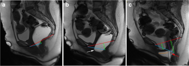
Pelvic floor dysfunctions represent a range of functional disorders that frequently occur in adult women, carrying a significant burden on the quality of life, and its incidence tends to increase attending to the expected aging of the population. Pelvic floor dysfunctions can manifest as incontinence, constipation, and prolapsed pelvic organs. Since pelvic floor weakness is frequently generalized and clinically underdiagnosed, imaging evaluation is of major importance, especially prior to surgical correction. Given some interobserver variability of soft-tissue measurements, MR defecography allows a noninvasive, radiation-free, multiplanar dynamic evaluation of the three pelvic compartments simultaneously and with high spatial and temporal resolution. Both static/anatomic and dynamic/functional findings are important, since pelvic disorders can manifest as whole pelvic floor weakness/dysfunction or as an isolated or single compartment disorder. Imaging has a preponderant role in accessing pelvic floor disorders, and dynamic MR defecography presents as a reliable option, being able to evaluate the entire pelvic floor for optimal patient management before surgery. The purpose of this article is to address the female pelvic anatomy and explain the appropriate MR Defecography protocol, along with all the anatomic points, lines, angles, and measurements needed for a correct interpretation, to later focus on the different disorders of the female pelvic floor, illustrated with MR defecography images, highlighting the role of this technique in accessing these pathologic conditions.

Keywords: Diagnostic imaging; Female pelvic floor; Magnetic resonance defecography.


Conflict of interest statement

Springer Nature remains neutral with regard to jurisdictional claims in published maps and institutional affiliations.

Figures

Fig. 1
Fig. 1
Normal female pelvic anatomy. Sagittal midline T2-TSE image demonstrating the perineal body (*) and the levator plate (white arrow). B, bladder; U, uterus; V, vagina; R, rectum
Fig. 2
Fig. 2
Pelvic compartments. Sagittal midline T2-TSE image representing the three pelvic compartments in different colors
Fig. 3
Fig. 3
Representative image of the pelvic floor ligaments and their relative anatomical positions in a female model
Fig. 4
Fig. 4
Pelvic muscles. (a, b) Coronal (a) and axial (b) T2-TSE images of the pelvic diaphragm, showing the iliococcygeus (white arrows) and puborectalis (red arrows) muscles
Fig. 5
Fig. 5
Pubococcygeal line (PCL). Sagittal midline T2-TSE image with the PCL represented (red line) connecting the inferior border of the pubic symphysis and the last coccygeal joint
Fig. 6
Fig. 6
Anorectal junction (ARJ). (a, b) Axial T2-TSE image (a) at the plane of the puborectalis muscle (red arrows), the same plane of the ARJ. Sagittal T2-TSE image (b) showing the posterior wall of the ARJ (white arrow)
Fig. 7
Fig. 7
Normal H and M lines. Sagittal midline T2-TSE image demonstrating the normal measurements of the H (4.47 cm) and M (1.16 cm) lines. Red line: PCL; blue line: H line; green line: M line
Fig. 8
Fig. 8
Normal anorectal angles. (a, b, c) Sagittal midline TrueFISP images at rest (a), during squeezing (b), and evacuation (c), with normal measurements of the ARA: 122° (a); 109° (b); 137° (c)
Fig. 9
Fig. 9
Importance of the evacuation phase. (a, b, c) Sagittal midline TrueFISP images at rest (a), during straining (b), and evacuation (c) of a patient with descending perineal syndrome with tricompartimental prolapse, anterior rectocele, and rectal mucosa intussusception. Note that, although in the straining phase (b) the H and M lines (blue and right green lines) are already above the normal values, indicating a descending perineal syndrome, and a mild bulge of the anterior rectal wall is seen (white arrow), only in the evacuation phase (c) the true pelvic floor disorder is adequately characterized, with a tricompartimental prolapse (cystocele, uterine prolapse, and rectal prolapse) the true size of the rectocele is documented (orange line: 4.8 cm) and the rectal mucosa intussusception is depicted (red arrow). Left green line: extent of cystocele; purple line: extent of uterine prolapse; black line: normal expected contour of the anterior anorectal wall; red line: PCL line
Fig. 10
Fig. 10
Moderate cystocele with urethral hypermobility. (a, b) Sagittal midline TrueFISP images at rest (a) and during evacuation (b), demonstrating descent of the bladder during evacuation (green line—3.8 cm) below the PCL (red line), as well as horizontalization of the urethral axis during defecation (white arrows). Note also an anterior rectocele in (b)
Fig. 11
Fig. 11
Uterine prolapse. Sagittal midline TrueFISP image at rest revealing an everted uterus filling of the vaginal space, which extends below the distal third of the vagina. Red line: PCL; purple line: extent of the uterine prolapse
Fig. 12
Fig. 12
Small rectoceles. Sagittal midline TrueFISP image during evacuation, showing both anterior and posterior small rectoceles. Black lines: normal expected contours of the anorectal wall; orange lines: extent of the rectoceles: 13 mm anterior and 9 mm posterior
Fig. 13
Fig. 13
Moderate anterior rectocele. (a, b) Sagittal midline TrueFISP images at rest (a) and during evacuation (b) revealing a moderate anterior rectocele (orange line: 37 mm). Note also a cystocele (green line) and a rectal intra-anal intussusception (white arrow). Red line: PCL line; black line: normal expected contour of the anterior anorectal wall
Fig. 14
Fig. 14
Rectal prolapse with a moderate uterine prolapse. (a, b, c) Sagittal midline TrueFISP images at the beginning (a), middle (b), and end (c) of the evacuation phase demonstrating a rectal intussusception extending beyond the anal sphincter (red arrows). Note also that it is accompanied by a moderate uterine prolapse and a peritoneocele, both aggravating along the different times of the evacuation phase. Red line: PCL; purple line: extent of the uterine prolapse
Fig. 15
Fig. 15
Peritoneocele with vaginal vault prolapse and rectal intussusception. (a, b) Sagittal midline TrueFISP images at rest (a) and during defecation (b) of a patient previously submitted to hysterectomy, showing a peritoneocele, where adipose peritoneal folds insinuate to the rectovaginal septum. It is associated with a prolapse of the vaginal vault, with the vaginal apex (white arrows) below the PCL (red line) during the evacuation. An apparent rectal invagination is also present (red arrow). Orange line: extent of the peritoneocele
Fig. 16
Fig. 16
Severe enterocele with vaginal vault prolapse, urethral hypermobility and urinary obstruction. (a, b) Sagittal midline TrueFISP images at rest (a) and during evacuation (b) of a patient previously submitted to hysterectomy, showing an enterocele. Note the herniation of small bowel loops into the rectovaginal septum. It is difficult to identify correctly the vaginal apex, but it is clear it is located below the PCL (red line), corresponding to a vaginal vault prolapse. The volume of the enterocele is so large that its mass effect over the vagina and urethra pushes them anteriorly, inducing also an horizontalization of their normal vertical axis—urethral hypermobility (white arrow)—and inducing obstruction of the bladder (the urinary volume of the bladder on both phases is similar, always above the PCL). Note also that the bladder is more distended than it should be. Although not relevant for this specific case, the bladder should be half distended, as preconized by the ESUR/ESGAR recommendations. Orange line: extent of the enterocele
Fig. 17
Fig. 17
Descending perineal syndrome with tricompartimental prolapse and anterior rectocele. (a, b) Sagittal midline TrueFISP images at rest (a) and during evacuation (b). H line (blue line) measures 5.6 cm (a) and 9.5 cm (b) and the M line (green line) measures 1.3 cm (a) and 4.7 cm (b), demonstrating widening and descent of the levator hiatus. Note the vertical orientation of the levator plate (white arrow). It is also associated with a tricompartimental prolapse and an anterior rectocele (*)
Fig. 18
Fig. 18
Descending perineal syndrome with tricompartimental prolapse. (a, b) Sagittal midline TruFISP images at rest (a) and during evacuation (b). H line (blue line) measures 7.7 cm (a) and 9.8 cm (b) and the M line (green line) measures 2.3 cm (a) and 4.5 cm (b), demonstrating widening and descent of the levator hiatus, even at rest but aggravating during defecation. It is also associated with tricompartimental prolapse, with a severe cystocele and a severe uterine everted prolapse. Note the verticalization of the levator plate (white arrow). Another interesting finding is that the rectal contrast is only partially expelled (b), because the mass effect by both the cystocele and uterine prolapse induces mechanical posterior obstruction (dilated rectum filled with ultrasound gel) causing constipation and incomplete evacuation (*). (c) Axial T2-TSE image at the pubic symphysis plane showing bilateral ureterohydronephosis causes by the cystocele (red arrows)
Fig. 19
Fig. 19
Puborectalis defect. Axial T2-TSE image of the puborectalis muscle, demonstrating a right posterior defect, asymmetry, and atrophy, with lipomatous involution (red arrow)
Fig. 20
Fig. 20
Descending perineal syndrome in a man. (a, b) Sagittal midline TrueFISP images at rest (a) and during evacuation (b) of a man with complaints of constipation and incomplete evacuation showing an increase of the H and M lines during evacuation phase (blue line, 88 mm; green line, 63 mm respectively), compatible with descending perineal syndrome with increased hiatal width and descent
Fig. 21
Fig. 21
Spastic pelvic floor syndrome. (a, b, c) Sagittal midline TrueFISP images of a 39-year-old patient at rest (a), during squeezing (b) and during evacuation (c). Note in (a) the ARA is 99° (elevated basal tonus), associated with the reduction of the ARA during defecation (c) with 50°, corresponding to a paradoxical contraction of the puborectalis muscle
Fig. 22
Fig. 22
Spastic pelvic floor syndrome. (a, b, c) Sagittal midline TrueFISP images of a man at rest (a), during squeezing (b) and evacuation (c), demonstrating a normal ARA angle at rest in (a) (118°), but revealing an increased reduction during squeezing in (b) (85°) and a lack of normal widening in the evacuation phase (c) (120°). During the dynamic evaluation, spastic contractions of the puborectalis muscle during evacuation were noted as well, and an incomplete evacuation with retention of ultrasound gel was present, all signs compatible with paradoxical contraction of the puborectalis muscle
Fig. 23
Fig. 23
Anal incontinence. (a, b) Sagittal midline TrueFISP images at rest (a) and during Valsava maneuver (b), with loss of the ultrasound gel in the second one. Notice the opening of the anal sphincter (red arrow). (c) Axial T2-TSE image of the internal anal sphincter demonstrating atrophy with lipomatous involution of the sphincter muscles (white arrow)

References

    1. García del Salto L, de Miguel Criado J, Aguilera del Hoyo LF et al (2014) MR imaging-based assessment of the female pelvic floor. Radiographics 34:1417–1439. - PubMed
    1. Olsen AL, Smith VJ, Bergstrom JO, Colling JC, Clark AL (1997) Epidemiology of surgically managed pelvic organ prolapse and urinary incontinence. Obstet Gynecol 89(4):501–506. - PubMed
    1. Luber KM, Boero S, Choe JY (2001) The demographics of pelvic floor disorders: current observations and future projections. Am J Obstet Gynecol 184(7):1496–1503. - PubMed
    1. Members of the Consensus Development Panel (1989) Consensus conference. Urinary incontinence in adults. JAMA 261(18):2685–2690. - PubMed
    1. Dolan LM, Hilton P (2010) Obstetric risk factors and pelvic floor dysfunction 20 years after first delivery. Int Urogynecol J 21:535–544. - PubMed

LinkOut - more resources