

Abdominal applications of ultrasound fusion imaging technique: liver, kidney, and pancreas
- PMID: 30689120
- PMCID: PMC6352389
- DOI: 10.1186/s13244-019-0692-z
Abdominal applications of ultrasound fusion imaging technique: liver, kidney, and pancreas
Abstract
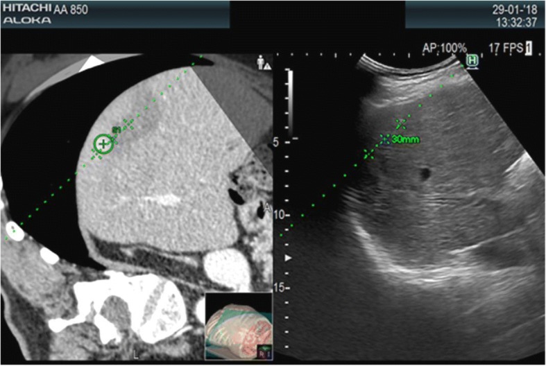
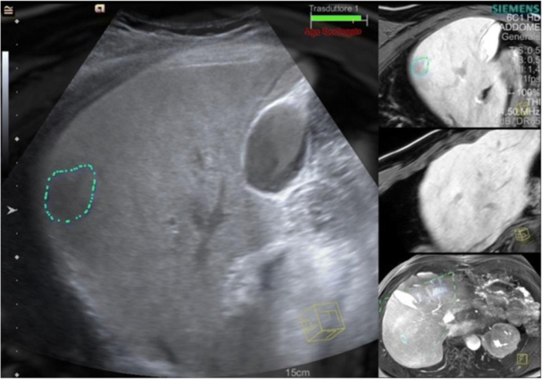
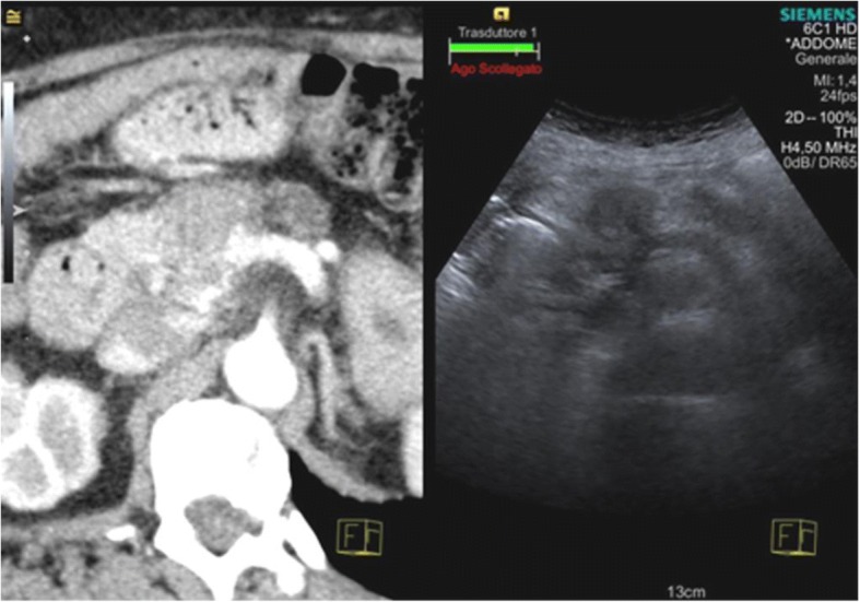
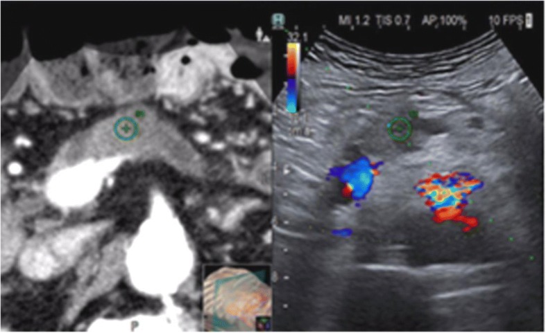
Fusion imaging allows exploitation of the strengths of all imaging modalities simultaneously, eliminating or minimizing the weaknesses of every single modality. Ultrasound (US) fusion imaging provides benefits in real time from both the dynamic information and spatial resolution of the normal US and the high-contrast resolution and wider field of view of the other imaging methods. US fusion imaging can also be associated with the use of different ultrasound techniques such as color Doppler US, elastography, and contrast-enhanced US (CEUS), for better localization and characterization of lesions. The present paper is focused on US fusion imaging technologies and clinical applications describing the possible use of this promising imaging technique in the liver, kidney, and pancreatic pathologies.
Keywords: Fusion imaging; Liver; Oncologic imaging; Tumor ablation; Ultrasound.
Conflict of interest statement
Springer Nature remains neutral with regard to jurisdictional claims in published maps and institutional affiliations.
Figures
References
-
- Lee MW, Lim HK, Kim YJ et al (2011) Percutaneous sonographically guided radio frequency ablation of hepatocellular carcinoma: causes of mistargeting and factors affecting the feasibility of a second ablation session. J Ultrasound Med 30(5):607–615. - PubMed
-
- Lorentzen T, Nolsøe CP, Ewertsen C et al (2015) EFSUMB guidelines on interventional ultrasound (INVUS), part I. general aspects (long version). Ultraschall Med 36(5):E1-14. - PubMed
-
- Song KD, Lee MW, Rhim H, Cha DI, Chong Y, Lim HK (2013) Fusion imaging-guided radiofrequency ablation for hepatocellular carcinomas not visible on conventional ultrasound. AJR Am J Roentgenol 201(5):1141–1147. - PubMed
LinkOut - more resources
Full Text Sources
