Review
. 2019 Apr 15;115(5):895-903.
doi: 10.1093/cvr/cvz013.

A current understanding of drug-induced QT prolongation and its implications for anticancer therapy

Affiliations
Review

A current understanding of drug-induced QT prolongation and its implications for anticancer therapy

Dan M Roden. Cardiovasc Res. .

Abstract

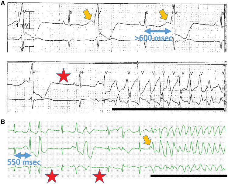
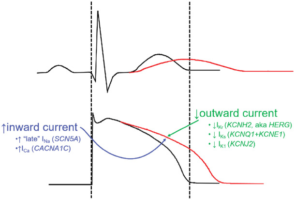
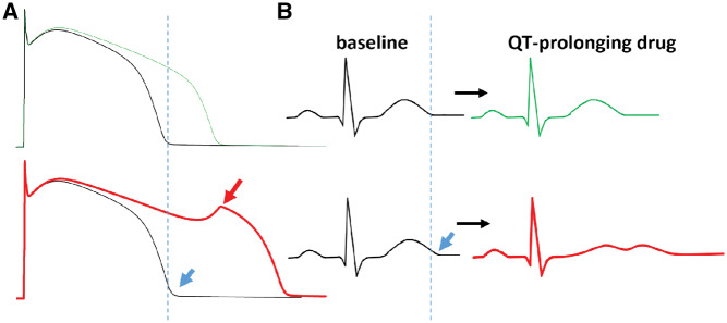
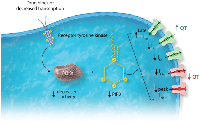
The QT interval, a global index of ventricular repolarization, varies among individuals and is influenced by diverse physiologic and pathophysiologic stimuli such as gender, age, heart rate, electrolyte concentrations, concomitant cardiac disease, and other diseases such as diabetes. Many drugs produce a small but reproducible effect on QT interval but in rare instances this is exaggerated and marked QT prolongation can provoke the polymorphic ventricular tachycardia 'torsades de pointes', which can cause syncope or sudden cardiac death. The generally accepted common mechanism whereby drugs prolong QT is block of a key repolarizing potassium current in heart, IKr, generated by expression of KCNH2, also known as HERG. Thus, evaluation of the potential that a new drug entity may cause torsades de pointes has relied on exposure of normal volunteers or patients to drug at usual and high concentrations, and on assessment of IKr block in vitro. More recent work, focusing on anticancer drugs with QT prolonging liability, is defining new pathways whereby drugs can prolong QT. Notably, the in vitro effects of some tyrosine kinase inhibitors to prolong cardiac action potentials (the cellular correlate of QT) can be rescued by intracellular phosphatidylinositol 3,4,5-trisphosphate, the downstream effector of phosphoinositide 3-kinase. This finding supports a role for inhibition of this enzyme, either directly or by inhibition of upstream kinases, to prolong QT through mechanisms that are being worked out, but include enhanced inward 'late' sodium current during the plateau of the action potential. The definition of non-IKr-dependent pathways to QT prolongation will be important for assessing risk, not only with anticancer therapies but also with other QT prolonging drugs and for generating a refined understanding how variable activity of intracellular signalling systems can modulate QT and associated arrhythmia risk.

Keywords: Arrhythmia; Cardio-oncology; PI3 Kinase; QT Prolongation; Torsades de pointes.


Figures

Figure 1
Figure 1
(A) Two rhythm strips obtained 5 min apart in a 25-year-old woman with sepsis, hypokalaemia, and therapy with ondansetron and a fluoroquinolone. The top rhythm strip shows ventricular bigeminy with a remarkable U wave (orange arrow) and prolonged repolarization. When the U wave amplitude is large, it is included in the QT measurement; in this case, the end of the QT-U complex is not seen and the QT-U exceeds 600 ms. The bottom strip shows a post-ectopic pause (red star), a sinus beat with a long QT-U, and an episode of the polymorphic ventricular tachycardia torsades de pointes (black line). QT interval was entirely normal without U waves after recovery. (B) Rhythm strip in a 60-year-old woman with Philadelphia chromosome positive acute leukaemia being treated with dasatinib and who developed cytokine storm when blinatumomab was added. The pauses, U wave, and TdP are indicated as above. QT interval was entirely normal pre-dasatinib, and prolonged to 500 ms with drug.
Figure 2
Figure 2
The normal QT interval (black, top; delineated by the dashed lines) is a rough indicator of the duration of action potentials in the ventricle (bottom). When the QT is prolonged (red), action potentials in at least some cells in the ventricle must be prolonged, and this can arise from increased inward current through sodium or potassium channels or decreased outward current through potassium channels. The genes encoding these key channels are also shown.
Figure 3
Figure 3
The concept of reduced repolarization reserve. (A) Computed (in silico) action potentials at baseline (black) are shown. The top panel is a normal action potential with the dashed line indicating the end of the action potential, and the bottom panel shows the effects of a 15% reduction in IKs, leading to a minimal prolongation of baseline action potential evident at the blue arrow. When IKr block is superimposed, the expected action potential prolongation (seen in the normal situation; green) is highly exaggerated in the presence of the minimal IKs lesion, and an early afterdepolarization is elicited (red arrow). (B) The effect on the electrocardiogram is shown. At baseline, the QT in the presence of the IKs lesion is minimally prolonged (blue arrow), but with drug exposure, there is minimal QT prolongation in the normal situation (top), and marked QT prolongation with development of a long, low-amplitude bifid T wave often seen in diLQTS.
Figure 4
Figure 4
Frequency of rare missense variants in congenital arrhythmia syndrome disease genes in patients with drug-induced long QT syndrome (diLQTS), drug-tolerant controls, and population controls from the Exome Sequencing Project. There was a high (∼50%) frequency of rare variants across all three groups, but far more patients with diLQTS had rare variants in arrhythmia genes encoding potassium channel subunits compared with the two control groups.
Figure 5
Figure 5
Distribution of genetic risk scores derived from a large genome-wide association study of baseline QT intervals. The distribution is significantly (P = 10−7) skewed to higher risk scores in subjects with drug-induced torsades de pointes (TdP cases) compared with drug exposed controls not developing TdP.
Figure 6
Figure 6
Role of phosphoinositide 3-kinase (PI3K) in ion channel signalling. As discussed in the text, occupancy of receptor tyrosine kinases (RTKs) activates PI3K (the α isoform in cardiomyocytes) which then generates its downstream effector phosphatidylinositol 3,4,5-trisphosphate (PIP3). Drug block of RTKs, or decreased transcription, results in enhanced late INa and decreased IKr and IKs, all of which (green) act to prolong action potential and QT, as well as decreased L-type calcium current and peak INa (orange) which would be expected to shorten QT. As described in the text, this effect on repolarization has now been reported with RTK inhibitors used in cancer, with other QT prolonging drugs formerly thought to act via IKr alone, and in diabetes. In each instance, the electrophysiologic abnormality is corrected by adding PIP3 to the intracellular solution.

References

    1. Schwartz PJ, Ackerman MJ, George AL Jr, Wilde AAM. Impact of genetics on the clinical management of channelopathies. J Am Coll Cardiol 2013;62:169–180. - PMC - PubMed
    1. Roden DM. Drug-induced prolongation of the QT Interval. N Engl J Med 2004;350:1013–1022. - PubMed
    1. Choy AM, Darbar D, Dell'Orto S, Roden DM. Exaggerated QT prolongation after cardioversion of atrial fibrillation. J Am Coll Cardiol 1999;34:396–401. - PubMed
    1. Astrom-Lilja C, Odeberg JM, Ekman E, Hagg S. Drug-induced torsades de pointes: a review of the Swedish pharmacovigilance database. Pharmacoepidemiol drug safety 2008;17:587–592. - PubMed
    1. Lazzerini PE, Laghi-Pasini F, Bertolozzi I, Morozzi G, Lorenzini S, Simpatico A, Selvi E, Bacarelli MR, Finizola F, Vanni F, Lazaro D, Aromolaran A, El Sherif N, Boutjdir M, Capecchi PL. Systemic inflammation as a novel QT-prolonging risk factor in patients with torsades de pointes. Heart 2017;103:1821–1829. - PubMed

Publication types

MeSH terms

Substances