Review
. 2019 Jun:56:78-86.
doi: 10.1016/j.sbi.2018.12.005. Epub 2019 Jan 26.

Protein O-fucosylation: structure and function

Affiliations
Review

Protein O-fucosylation: structure and function

Bernadette C Holdener et al. Curr Opin Struct Biol. 2019 Jun.

Abstract

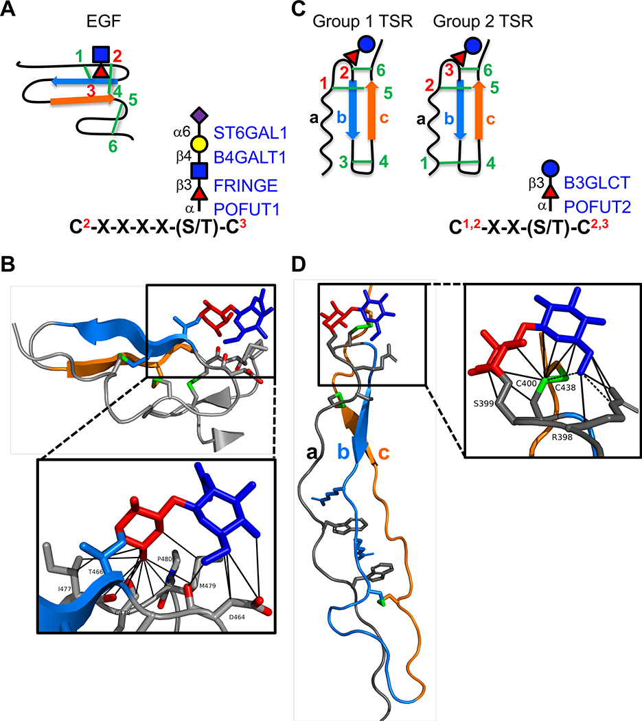
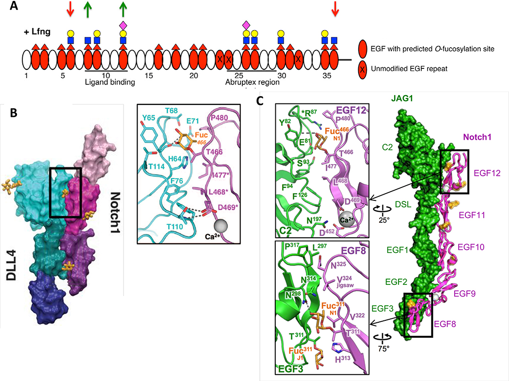
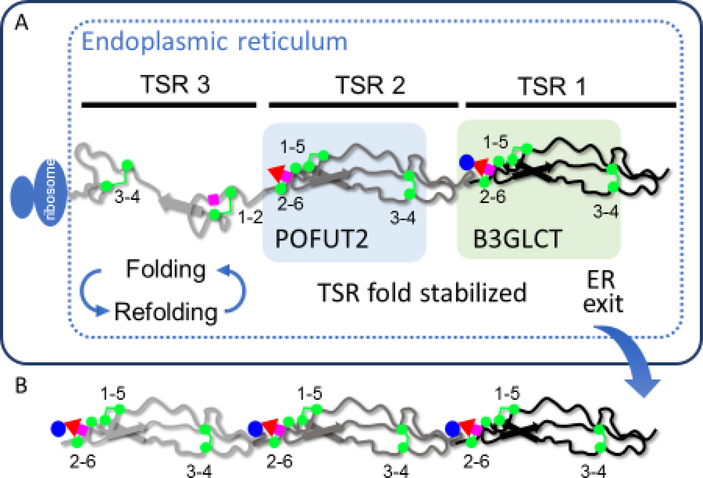
Fucose is a common terminal modification on protein and lipid glycans. Fucose can also be directly linked to protein via an O-linkage to Serine or Threonine residues located within consensus sequences contained in Epidermal Growth Factor-like (EGF) repeats and Thrombospondin Type 1 Repeats (TSRs). In this context, fucose is added exclusively to properly folded EGF repeats and TSRs by Protein O-fucosyltransferases 1 and 2, respectively. In both cases, the O-linked fucose can also be elongated with other sugars. Here, we describe the biological importance of these O-fucose glycans and molecular mechanisms by which they affect the function of the proteins they modify. O-Fucosylation of EGF repeats modulates the Notch signaling pathway, while O-fucosylation of TSRs is predicted to influence secretion of targets including several extracellular proteases. Recent data show O-fucose glycans mediate their effects by participating in both intermolecular and intramolecular interactions.


Conflict of interest statement

Conflict of interest: The authors declare no conflict of interest.

Figures

Figure 1.
Figure 1.. EGF repeats and TSRs are modified by O-fucose glycans A.
(left) Cartoon showing disulfide bonding pattern (green lines) in an EGF repeat. Beta strands are indicated by blue and orange arrows. Site of O-fucosylation and GlcNAc elongation are indicated by red triangle and blue square, respectively. (right) Consensus sequence for POFUT1 modification. C2 and C3 are the second and third conserved cysteine in the EGF repeat. Enzymes responsible for addition of each sugar are indicated in blue on the right with linkages in black on the left. Fucose, red triangle; GlcNAc, blue square; Galactose, yellow circle; Sialic Acid, purple diamond. B. Structure of NOTCH1 EGF12 modified with a GlcNAcβ1–3Fucose disaccharide (from PDB ID 4D0E). Beta strands colored as in A. Fucose in red, GlcNAc in blue, disulfide bonds in green, oxygen atoms highlighted in red. Box shows zoomed in region highlighting interactions of the disaccharide with underlying amino acids identified by MolProbity [73,74] (van der Waals, solid lines). Structures rendered in PyMOL (Version 2.2.2). C. (left) Cartoons showing the two distinct disulfide bonding patterns for TSRs. Beta strands are indicated by blue and orange arrows. Position of O-fucosylation and elongation with glucose are indicated by red triangle and blue circle, respectively. (right) Consensus sequence for POFUT2 modification. The C’s can be C1 and C2 or C2 and C3 depending on whether the TSR is Group 1 or Group 2. Enzymes responsible for addition of each sugar are indicated in blue on the right with linkages in black on the left. Fucose, red triangle; Glucose, blue circle. D. Structure of ADAMTS13 TSR1 modified with Glucoseβ1–3-Fucose disaccharide (from PDB ID 3GHM). The three strands (a, b, and c) of the TSRs are color coded the same in the cartoons (C) and the structure. Fucose in red, glucose in blue, disulfide bonds in green. Box shows zoomed in region highlighting interactions of the disaccharide with underlying amino acids identified by MolProbity [73,74] (H-bonds, dashed lines; van der Waals, solid lines). Structures rendered in PyMOL (Version 2.2.2).
Figure 2.
Figure 2.. O-Fucose on EGF8 and EGF12 of NOTCH1 is in direct contact with ligands.
A. Domain map of mouse NOTCH1 EGF1–36 showing which EGF repeats are modified by O-fucose and elongated by LFNG (modified from [26]). MFNG elongates similarly, but RFNG only modifies O-fucose on EGF8, 12 and 26. Note that Fringe enzymes were overexpressed in a Fringe-deficient background in these studies. Down red arrows indicate sites where Fringe modification inhibits JAG1-NOTCH1 activation. Green up arrows indicate sites where Fringe modification enhances DLL1-NOTCH1 activation. Fucose, red triangle; GlcNAc, blue square; Galactose, yellow circle; Sialic Acid, purple diamond. B. Co-crystal structure of NOTCH1 EGF11–13 (shades of magenta/purple) and DLL4 (N-terminus to EGF3, shades of blue/green) (modified from [52]). Inset shows direct interaction between O-fucose on NOTCH1 EGF12 with residues in DLL4. C. Co-crystal structure of NOTCH1 EGF8–12 and JAG1 N-EGF3 (modified from [53]). Inset shows direct contacts between O-fucose on EGF8 and EGF12 and residues in JAG1. Note that the structures in B and C were obtained after directed evolution of the ligands toward stronger affinities.
Figure 3.
Figure 3.. O-Fucosylation stabilizes TSRs by interacting with underlying amino acids.
A. A hypothetical protein with 3 TSRs is being translated and is folding in the ER. TSR3 is in the folding cycle, with some correct and some incorrect disulfide bonds. TSR3 is not modified by O-fucose glycans. TSR2 is fully folded, stabilized by addition of fucose by POFUT2, and is no longer part of a folding cycle. TSR1 is further stabilized by addition of glucose by B3GLCT. Note that the folding pathway for a TSR is not known, so this pathway is hypothetical. B. Once all TSRs with a POFUT2 consensus sequence are modified with Glucoseβ1–3Fucose disaccharide and the protein is fully folded, the native protein exits the ER, transits the secretory system, and is either directed to the cell surface or secreted from the cell depending upon the target protein properties.

References

    1. Schneider M, Al-Shareffi E, Haltiwanger RS: Biological functions of fucose in mammals. Glycobiology 2017, 27:601–618. - PMC - PubMed
    1. Varshney S, Stanley P: Multiple Roles for O-Glycans in Notch Signalling. FEBS Lett 2018. 592:3819–3834 Excellent, recent comprehensive review of O-glycans occuring on the Notch receptor - PMC - PubMed
    1. Haltom AR, Jafar-Nejad H: The multiple roles of epidermal growth factor repeat O-glycans in animal development. Glycobiology 2015, 25:1027–1042. - PMC - PubMed
    1. Vasudevan D, Haltiwanger RS: Novel roles for O-linked glycans in protein folding. Glycoconj J 2014, 31:417–426. - PMC - PubMed
    1. Swearingen KE, Lindner SE, Shi L, Shears MJ, Harupa A, Hopp CS, Vaughan AM, Springer TA, Moritz RL, Kappe SH, et al.: Interrogating the Plasmodium Sporozoite Surface: Identification of Surface-Exposed Proteins and Demonstration of Glycosylation on CSP and TRAP by Mass Spectrometry-Based Proteomics. PLoS Pathog 2016, 12:e1005606. - PMC - PubMed

Publication types

LinkOut - more resources