. 2019 Mar 1:14:364-376.
doi: 10.1016/j.omtn.2018.12.008. Epub 2018 Dec 20.

Viral Vector-Based Delivery of CRISPR/Cas9 and Donor DNA for Homology-Directed Repair in an In Vitro Model for Canine Hemophilia B

Affiliations

Viral Vector-Based Delivery of CRISPR/Cas9 and Donor DNA for Homology-Directed Repair in an In Vitro Model for Canine Hemophilia B

Jian Gao et al. Mol Ther Nucleic Acids. .

Abstract

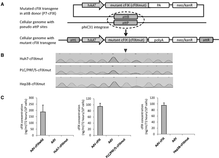
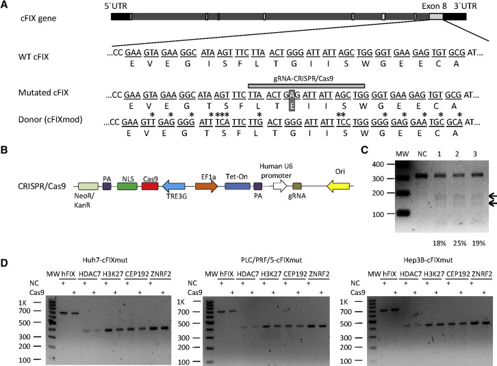
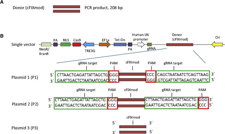
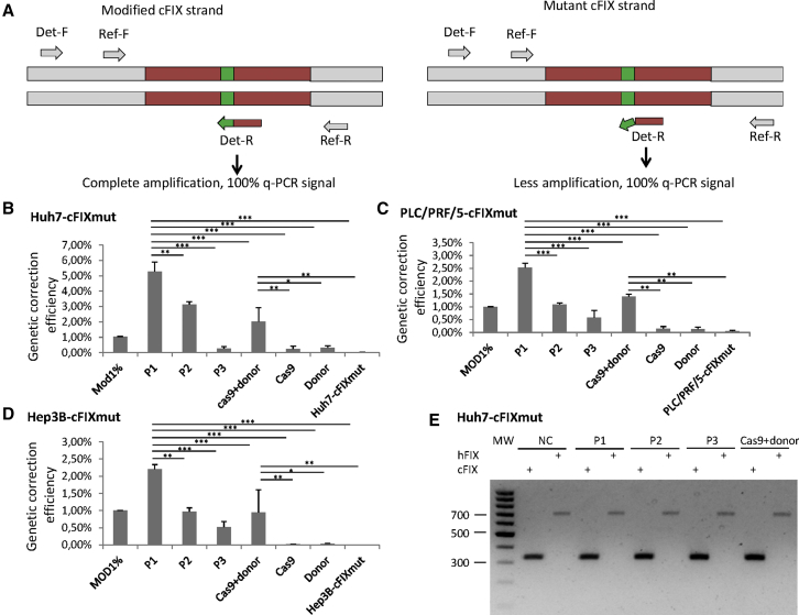
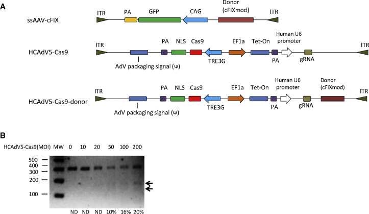
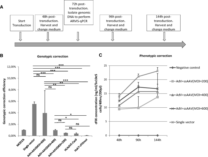
Gene therapy represents an attractive alternative to treat hemophilia B. Here we established three hepatocyte-derived cell lines based on Huh7, PLC/PRF/5, and Hep3B cells stably carrying a mutated canine FIX (cFIXmut) transgene containing a single point mutation in the catalytic domain. Based on these in vitro models resembling a commonly used canine large animal model, the tetracycline-controlled transcriptional activator (Tet-on)-inducible CRISPR/Cas9 system and an optimized donor were used to correct mutated cFIX gene through homology-directed repair (HDR). For efficient delivery of designer nuclease and donor DNA, we produced a high-capacity adenovirus vector type 5 (HCAdV5) containing the Tet-on-inducible cFIX-specific CRISPR/Cas9 system and a single-stranded adeno-associated virus type 2 vector (ssAAV2) containing the modified donor. Moreover, we designed a single HCAdV5 delivering all components for HDR. Our amplification-refractory mutation system based on qPCR analysis (ARMS-qPCR) revealed that the single vector application in Huh7-cFIXmut cells resulted in up to 5.52% HDR efficiencies, which was superior to the two-vector strategy. Furthermore the single vector also resulted in increased phenotypic correction efficiencies assayed by ELISA. We conclude that HDR in combination with viral vector delivery holds great promise for the correction of mutated FIX in disease models.

Keywords: CRISPR/Cas9; HCAdV5; gene therapy; hemophilia B; homology directed repair; single vector; ssAAV2.


Figures

Figure 1
Figure 1
Construction of Stable Mutated Liver Cells Using phiC31 Integrase (A) The vector containing the attB site, neo/kanR, and the mutated canine factor IX (cFIX) expression cassette and the phiC31 expression plasmid were co-transfected into liver-derived cell lines (Huh7 cells, PLC/PRF/5 cells, and Hep3B cells). In the presence of phiC31 integrase, an attB-containing donor can be unidirectionally integrated into a target cell genome, resulting in an integrated transgene flanked by attL and attRR sites. After selection with G418 for 3 weeks and sequence verification, the stable mutated cFIX cells (Huh7-cFIXmut, PLC/PRF/5-cFIXmut, and Hep3B-cFIXmut) were prepared for homology-directed repair (HDR) correction experiments. hAAT, human alpha-1-antitrypsin promoter; PA, polyadenylation signal; neo, neomycin; kanR, kanamycin resistance. (B) The cFIX fragment of stable cell clones was checked using Sanger sequencing. The black arrow indicates the point mutation G1477A in cFIX. (C) The supernatants of cFIX stable cell clones were checked using ELISA. Data points represent SEM of three independent experiments performed in triplicates. AdV-cFIX, supernatant of cells infected with adenovirus vector carrying a wild cFIX transgene; AdV, supernatant of cells infected with an irrelevant adenovirus vector; Huh7-cFIXmut, PLC/PRF/5-cFIXmut, and Hep3B-cFIXmut, the supernatants of the respective cFIX stable cell clones.
Figure 2
Figure 2
Construction of Non-viral Homology-Directed System for Correcting Mutated Canine Coagulation Factor IX (A) Schematic outline of the cFIX target locus. The top panel shows the cFIX gene, including the UTRs (black bars), introns (bright gray bars), and exons (light gray bars). Nucleotide and amino acid sequences of wild-type cFIX (WT cFIX) and a major disease causing cFIX mutation (G1477A, mutated cFIX) are shown (white letters in dark square). The bottom panel shows the mutated donor sequence (cFIXmod) used in this study (mutations are marked with an asterisk), which results in the identical amino acid sequence as WT cFIX. The gray horizontal bar schematically shows the guide RNA (gRNA)-binding site used in this study (gRNA-CRISPR/Cas9). Differences in the donor and mutated cFIX sequences are marked with stars. (B) Schematic outline of the DNA sequences contained in the Tet-on-inducible CRISPR/Cas9 for cutting the cFIX-mutated strand and the donor DNA that was transfected as PCR product (cFIXmod). NLS, nuclear localization signal; PA, polyadenylation signal; TREG3, TRE3G promoter; EF1a, human elongation factor-1 alpha promoter; gRNA, guide RNA; Tet-on, tetracycline-controlled transcriptional activation. (C) CRISPR/Cas9 nuclease activity measured by T7E1 assay after transfection with the CRISPR/Cas9-encoding plasmid and the donor DNA. MW, molecular-weight size marker; NC, negative control referring to the mixture of untreated Huh7-cFIXmut, PLC/PRF/5-cFIXmut, and Hep3B-cFIXmut cells; 1, CRISPR/Cas9-treated Huh7-cFIXmut cells; 2, CRISPR/Cas9-treated PLC/PRF/5-cFIXmut cells; 3, CRISPR/Cas9-treated Hep3B-cFIXmut cells. (D) CRISPR/Cas9 nuclease off-target analysis of the top 5 predicted off-target sites. These were the histone deacetylase 7 gene (HDAC7), DNA-packaging protein Histone H3 (H3K27), the centrosomal protein of 192 kDa (CEP192), and the Zinc and Ring Finger 2 gene (ZNRF2), which were analyzed in Huh7-cFIXmut, PLC/PRF/5-cFIXmut, and Hep3B-cFIXmut cells by T7E1 assay after transfection with the CRISPR/Cas9-encoding plasmid.
Figure 3
Figure 3
Design of Donor DNA Sequences for the Two-Vector and the Single-Vector Systems (A) Donor DNA used for the two-vector system. A PCR product of 208 bp (cFIXmod) covering the mutated cFIX location was used. (B) Schematic diagram of single vectors containing the Tet-on-inducible CRISPR/Cas9 for cutting the cFIX-mutated strand and the donor DNA. Three molecular setups of single vectors, which delivered all components for HDR, were designed: plasmid 1 (P1), plasmid 2 (P2), and plasmid 3 (P3). For P1 and P2, the donor is flanked by the gRNA recognition sequence. Note that the gRNA recognition sequences in P1 are in the opposite orientation while the gRNA recognition sequences of P2 flank the donor DNA in the identical orientation. The donor contained in plasmid P3 lacks the gRNA recognition sequence. NLS, nuclear localization signal; PA, polyadenylation signal; TREG3, TRE3G promoter; EF1a, human elongation factor-1 alpha promoter; gRNA, guide RNA; Tet-on, tetracycline-controlled transcriptional activator.
Figure 4
Figure 4
Gene Correction of Mutated cFIX through Naked DNA (A) The principle of the amplification-refractory mutation system qPCR (ARMS-qPCR) assay. Two qPCRs of each sample are performed. Primers of the reference PCR (Re-F,-R) amplify the upstream area of the HR region. The forward detection primer (Det-F) binds the upstream area of the HR region, and the reverse primer (Det-R) specifically binds to the HR cassette. (B–D) Naked DNA-mediated HDR events in mutated cFIX stable cell lines measured via ARMS-qPCR. Mod1%, Cas9 + donor, Cas9, Donor, and cell-cFIXmut display controls containing 1% modified template, cells transfected with CRISPR/Cas9 and optimized donor sequence, cells transfected with CRISPR/Cas9, cells transfected with optimized donor sequence, and cFIX stable cell lines Huh7-cFIXmut (B), PLC-PRF-5-cFIXmut (C), and Hep3B-cFICmut (D). (E) Indels measured by T7E1 assay after transfection with the non-viral homology-directed repair plasmids at the endogenous hFIX locus and the uncorrected cFIX transgene in Huh7-cFIXmut cells. MW, molecular weight markers; NC, negative control, PLC-cFIXmut or Hep3B-cFIXmut without treatment. P1, P2, P3, and Cas9 + donor display cells treated with plasmid 1, plasmid 2, plasmid 3, or Cas9 + donor. Data points represent SEM of three independent experiments performed in triplicates. *p < 0.05, **p < 0.01, ***p < 0.001.
Figure 5
Figure 5
Gene Correction of Mutated cFIX through the Viral Vectors HCAdV5-Cas9 and ssAAV2-cFIX (A) Schematic outline of HCAdV5-Cas9 and ssAAV2-cFIX genomes. ITR, inverted terminal repeat; NLS, nuclear localization signal; PA, polyadenylation signal; TREG3, TRE3G promoter; EF1a, human elongation factor-1 alpha promoter; CAG, cytomegalovirus (CMV) enhancer fused to the chicken beta-actin promoter; gRNA, guide RNA; Tet-on, tetracycline-controlled transcriptional activation. (B) HCAdV5-Cas9 nuclease activity measured by T7E1 assay. MW, molecular-weight size marker; ND, no detection.
Figure 6
Figure 6
Genotypic and Phenotypic Corrections in Huh7-cFIXmut Cells after Co-infection with the Gene Correction Vectors HCAdV5-Cas9-cFIX or HCAdV5-Cas9 and ssAAV2-cFIX (A) Schematic outline of the experimental setup. Cellular genomic DNA was extracted at 72 h post-transduction for ARMS-qPCR assay, and cell media were collected at 48, 96, and 144 h post-transduction for the ELISA. (B) HCAdV5-Cas9-cFIX- or HCAdV5-Cas9- and ssAAV2-cFIX-mediated HDR events in mutated cFIX stable cells measured via ARMS-qPCR. Mod1%, controls containing 1% modified template; single vector, Huh7-cFIXmut cells transduced with HCAdV5-Cas9-cFIX; AdV + AAV (MOI 200), AdV + AAV (MOI 400),and AdV + AAV (MOI 600), Huh7-cFIXmut cells transduced with HCAdV5-Cas9 and ssAAV2-cFIX of different MOIs; Huh7-cFIXmut cells, mutated cFIX stable Huh7 cells; HCAdV5-Cas9, Huh7-cFIXmut cells transduced with HCAdV5-Cas9. (C) ELISA of cFIX concentration in the supernatant of Huh7-cFIXmut cells infected with HCAdV5-Cas9-cFIX or HCAdV-Cas9 and MOIs 200, 400, and 600 for ssAAV-cFIX. Data points represent SEM of three independent experiments performed in triplicates. *p < 0.05, **p < 0.01, ***p < 0.001.

Similar articles

Cited by

References

    1. Nathwani A.C., Tuddenham E.G. Epidemiology of coagulation disorders. Baillieres Clin. Haematol. 1992;5:383–439. - PubMed
    1. Sun J., Hua B., Livingston E.W., Taves S., Johansen P.B., Hoffman M., Ezban M., Monroe D.M., Bateman T.A., Monahan P.E. Abnormal joint and bone wound healing in hemophilia mice is improved by extending factor IX activity after hemarthrosis. Blood. 2017;129:2161–2171. - PMC - PubMed
    1. Kay M.A., Rothenberg S., Landen C.N., Bellinger D.A., Leland F., Toman C., Finegold M., Thompson A.R., Read M.S., Brinkhous K.M. In vivo gene therapy of hemophilia B: sustained partial correction in factor IX-deficient dogs. Science. 1993;262:117–119. - PubMed
    1. Kay M.A., Manno C.S., Ragni M.V., Larson P.J., Couto L.B., McClelland A., Glader B., Chew A.J., Tai S.J., Herzog R.W. Evidence for gene transfer and expression of factor IX in haemophilia B patients treated with an AAV vector. Nat. Genet. 2000;24:257–261. - PubMed
    1. Nathwani A.C., Tuddenham E.G., Rangarajan S., Rosales C., McIntosh J., Linch D.C., Chowdary P., Riddell A., Pie A.J., Harrington C. Adenovirus-associated virus vector-mediated gene transfer in hemophilia B. N. Engl. J. Med. 2011;365:2357–2365. - PMC - PubMed

LinkOut - more resources