Review
. 2019 Mar:197:345-367.
doi: 10.1016/j.biomaterials.2019.01.011. Epub 2019 Jan 7.

Ocular adhesives: Design, chemistry, crosslinking mechanisms, and applications

Affiliations
Review

Ocular adhesives: Design, chemistry, crosslinking mechanisms, and applications

Grissel Trujillo-de Santiago et al. Biomaterials. 2019 Mar.

Abstract

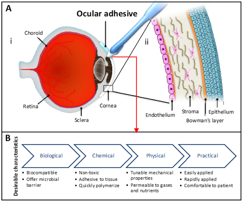
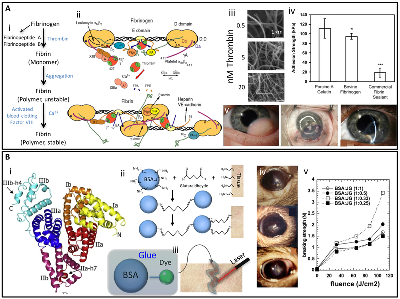
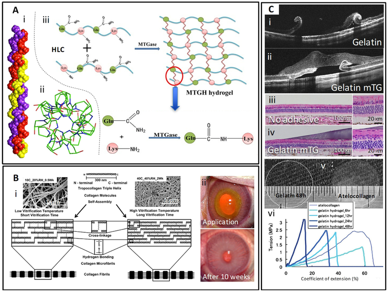
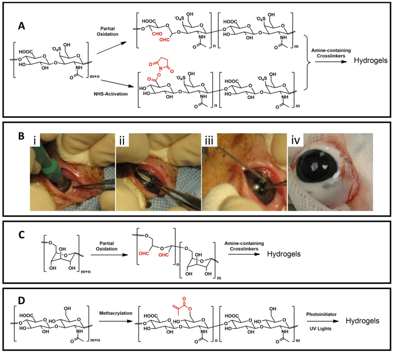
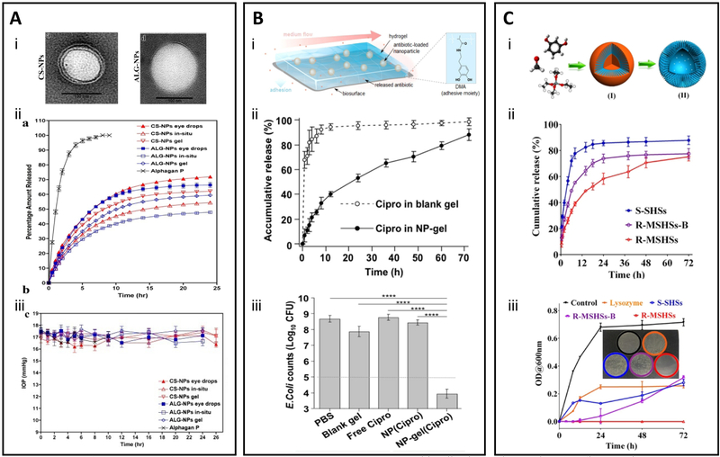
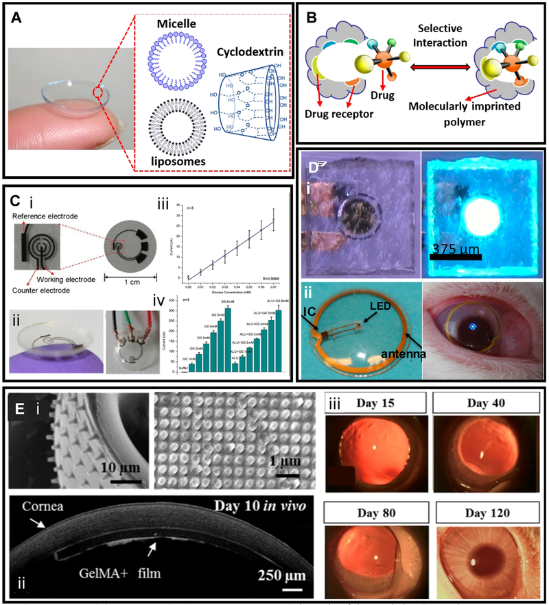
Closure of ocular wounds after an accident or surgery is typically performed by suturing, which is associated with numerous potential complications, including suture breakage, inflammation, secondary neovascularization, erosion to the surface and secondary infection, and astigmatism; for example, more than half of post-corneal transplant infections are due to suture related complications. Tissue adhesives provide promising substitutes for sutures in ophthalmic surgery. Ocular adhesives are not only intended to address the shortcomings of sutures, but also designed to be easy to use, and can potentially minimize post-operative complications. Herein, recent progress in the design, synthesis, and application of ocular adhesives, along with their advantages, limitations, and potential are discussed. This review covers two main classes of ocular adhesives: (1) synthetic adhesives based on cyanoacrylates, polyethylene glycol (PEG), and other synthetic polymers, and (2) adhesives based on naturally derived polymers, such as proteins and polysaccharides. In addition, different technologies to cover and protect ocular wounds such as contact bandage lenses, contact lenses coupled with novel technologies, and decellularized corneas are discussed. Continued advances in this area can help improve both patient satisfaction and clinical outcomes.

Keywords: Bioadhesives and sealants; Drug delivery; Natural and synthetic; Ocular.


Figures

Figure 1.
Figure 1.. Desirable characteristics in an ocular adhesive and eye anatomy and cornea structure.
(A) Anatomy of (i) eye and (ii) cornea. (B) Biological, chemical, physical, and practical characteristics that an ideal ocular adhesive should exhibit.
Figure 2.
Figure 2.. Structure, properties and clinical applications of cyanoacrylate adhesives.
(A) Preparation and anionic polymerization reaction (crosslinking) of cyanoacrylate monomers. Adapted from Scognamiglio et al. [61] with permission from Wiley, copyright 2016. (B) Cyanoacrylate glue applications techniques, (i) traditional technique: direct application of a drop of cyanoacrylate glue on a pre-dried ocular tissue, and (ii) patient eye treated with this technique; (iii) infant cannula technique: a small disc of a non-adhesive material is cut with a punch biopsy, an infant cannula (without needle) is used to pick up and hold the disc, a drop of glue is applied on the disc, and finally the glue is placed gently on the pre-dried ocular tissue, (iv) patient eye treated with this technique, (v) corneal tissue repaired with this technique. Adapted from Rana et al. [50] with permission from Elsevier, copyright 2013. (C) A polyglactin mesh glued with cyanoacrylate on a cadaver eye for strabismus surgery application, and effect of the polymerization time and surface area on the bonding strength of the mesh to the eye tissue. Adapted from Bona et al. [60] with permission from Elsevier, copyright 2014.
Figure 3.
Figure 3.. Molecular structure and properties of PEG-based adhesives.
(A) PEGDA synthesis (i) PEG functionalized with diacrylated groups formed a 3D hydrogel network in the presence of a photoinitiator upon exposure to light. Physical and biochemical properties of resulting hydrogels were customized by modifying hydrogel formulations. (ii) Effect of PEGDA molecular weight on water swelling behavior and on pattern resolution on molded PEGDA hydrogels. (iii) Effect of PEGDA molecular weight on non-specific cell attachment. Adapted from Yanez-Soto et al. [72] with permission from Wiley, copyright 2013. (B) (i) Chemical modifications to functionalize PEGDA hydrogels with RGD peptide motifs, Micro-molded PEGDA hydrogels (ii) with and (iii) without RGD functionalization to tune cell attachment and to direct the alignment of human coronal epithelial cells. Scale bar: 100 μm. Adapted from Yanez-Soto et al. [72] with permission from Wiley, copyright 2013. (C) (i) Scheme of the sealing of scleral incisions using PEG adhesive, (ii) Photographic images of sealing of a scleral incision in a rabbit vitrectomy model with PEG adhesive, (iii) Photographs of sectioned samples of a sclerotomy site closed with the PEG-based adhesive (the conjunctival incision is indicated by the arrow). Excessive inflammation is not evident. (Scale bar: 500 μm). Adapted from Hoshi et al. [81] with permission from Association for Research in Vision and Ophthalmology, copyright 2016.
Figure 4.
Figure 4.. Structure, properties, and applications of dendritic adhesives.
(A) Schematic illustration of different generations of dendritic adhesives from G0 to G4, which were prepared via divergent synthesis. The central core, inner branches, and peripheral groups were three main structural regions of these macromolecules. Adapted from Grinstaff et al. [99] with permission from Wiley, copyright 2002. (B) A method to apply and crosslink a ([G1]-PGLSAMA)2-PEG dendritic adhesive. An incision of 4.1 mm was made on enucleated an eye cornea, 15–20 μL of adhesive was placed on the surface of the pre-dried wound, the adhesive was photo-crosslinked using argon laser (diffuse beam; 200 mW; 1 sec), the sealed wound withstood an IOP of 109.6 ± 82.7 mmHg before leakage. Adapted from Velazquez et al. [98] with permission from American Medical Association, copyright 2004. (C) Images of histology transversal cuts of chicken cornea wounds treated with a ([G1]-PGLSAMA)2-PEG dendritic adhesive and suture after 28 days. Adapted from Grinstaff et al. [2] with permission from Elsevier, copyright 2007. (D) Dendrimers used as crosslinking agents of collagen hydrogels. (i) Mechanical properties and (ii) cell growth of collagen hydrogel crosslinked with generation 2 polypropyleneimine octaamine dendrimer compared to collagen hydrogels crosslinked with 1-ethyl-3-(3-dimethyl aminopropyl) carbodiimide hydrochloride and glutaraldehyde. Adapted from Duan and Sheardown et al. [97] with permission from Elsevier, copyright 2006.
Figure 5.
Figure 5.. Structure, properties, and applications of fibrin- and albumin-based adhiesives.
(A) Mechanism of fibrin clot formation: (i) Schematic representation of the conversion from fibrinogen to fibrin and subsequent polymerization/crosslinking mechanisms [133], (ii) Fibrinogen structure and its thrombin-mediated conversion to fibrin. Binding sites for the main molecular actors that participate in fibrinogen functions are illustrated. Adapted from Mosesson et al. [109] with permission from Wiley, copyright 2005, (iii) Fibrin clots resulting from the addition of thrombin (0.5–20 nM) solutions to fibrinogen (2 mg/ mL) as observed by SEM. (Scale bar: 1 μm). Adapted from Wolberg et al. [110] with permission from Elsevier, copyright 2007, (iv) Comparison of adhesive strengths of photocrosslinked porcine gelatin, photocrosslinked bovine fibrinogen, and a commercial fibrin tissue sealant (Tisseel). Adapted from Elvin et al. [137] with permission from Elsevier, copyright 2010, (v) An application of fibrin glue in ophthalmology. Left eye of an infant patient with an inferotemporal growth before surgery, graft after attachment with fibrin adhesive, and 10 weeks later. Adequate graft integration with no edema and minimum haze was observed. Adapted from Zhou et al. [118] with permission from Healio, copyright 2016; (B) Albumin based adhesives: (i) The 3D structure of albumin; a globular protein abundantly present in animal serum, (ii) Mechanism of crosslinking and tissue adhesion of BioGlue®, (iii) Schematic illustration of laser soldering using albumin-based solders. Adapted from Chao et al. [126] with permission from Wiley, copyright 2003, (iv) Rat eyes glued with albumin soldering after corneal epithelium removal surgery, and (v) ex-vivo breaking strength measure on mouse skin soldered with albumin-based solder. The ratio of protein to fluorescent dye has a significant effect on breaking strength as measured by tensiometer. Adapted from Khadem et al. [131] with permission from Wiley, copyright 2004.
Figure 6.
Figure 6.. Structure, properties, and applications of collagen and gelatin-based adhesives.
(A) Collagen-based adhesives. (i) 3D structure of collagen; collagen triple helix with sequence (POG)10, (ii) The top view of helical twist in the collagen structure. The top view of the T3–785 peptide (crystal structure), represents the first three POG triplets on each chain. Adapted from Bella [138] with permission from Portland Press, copyright 2016, (iii) Schematic representation of the mechanism of crosslinking of collagen (or gelatin) in the presence of transglutaminase. Transglutaminase enzyme forms amide bonds between the acyl groups in glutamine and the amino groups in lysine present in the protein (collagen or gelatin) chains. Adapted from Zhao et al. [152] with permission from Elsevier, copyright 2016; (B) Collagen and vitrigels. (i) Schematic explanation of the effect of vitrification temperature and time on the microstructure of collagen vitrigels. Insets are SEM images of the corresponding collagen vitrigels. Adapted from Calderón-Colón et al. [151] with permission from Elsevier, copyright 2012, (ii) Rabbit eye model with stromal wound treated with collagen vitrigel membrane and fibrin glue. Adapted from Chae et al. [163] with permission from Wiley, copyright 2015. (C) Gelatin-based adhesives. Optical coherence tomography (OCT) of a rabbit eye with retinal detachment treated (i) with gelatin or (ii) gelatin crosslinked with transglutaminase as observed 3 days of application. Images of histology transversal cuts of (iii) untreated and (iv) treated eyes three days after treatment. Gelatin-mTG adhesives tightly adhered to the retinal surface. Adapted from Yamamoto et al. [164] with permission from Springer, copyright 2013, (v) Transparency of gelatin and atelocollagen films after under wet conditions, (vi) Strain vs stress curves for different gelatin hydrogel films. Adapted from Watanabe et al. [170] with permission from Mary Ann Liebert, copyright 2011.
Figure 7.
Figure 7.. Examples of polysaccharide-based adhesives.
(A) Commonly used chemical crosslinking mechanisms for chondroitin sulfate-based hydrogels. (B) Application of NHS-modified chondroitin sulfate/amine PEG sealants in a swine eye model, (i) A 6.0 mm defect was made in the cornea with a trephine, (ii) an incision was made to get a flap, (iii) the sealant was applied to glue the flap to the stroma, (iv) the flap was tightly adhered 2 weeks after surgery. Adapted from Strehin et al. [173] with permission from Elsevier, copyright 2009. (C) Schematic illustration of the partial oxidation of dextran and the formatting mechanism of hydrogels crosslinked by amine-containing crosslinkers. (D) Schematic illustration of the methacrylation and photocrosslinking reactions of hyaluronic acid-based sealant.
Figure 8.
Figure 8.. Examples of bioadhesives used for drug delivery.
(A) A bioadhesive to lower the IOP. (i) TEM images of the brimonidine-loaded chitosan (CS) and alginate (ALG) nanoparticles, (ii) Release profile of brimonidine from the nanoparticles eye drops, gel and in situ, (iii) IOP effect of the formulation. Adapted from Ibrahim et al. [193] with permission from ARVO, copyright 2015; (B) (i) Schematic of the NP-gel system, consisting of ciprofloxacin-loaded nanoparticles in a 3D adhesive hydrogel, (ii) Release profile of ciprofloxacin in vitro, (iii) Effect of the adhesive on E. coli bacterial film in vivo. Adapted from Zhang et al. [194] with permission from American Chemical Society, copyright 2016; (C) (i) Schematic of the hollow nanocarriers loaded with lysozyme, with rough surface to enhance adhesion to bacteria, (ii) Release profile of lysozyme in vitro for smooth silica hollow spheres (S-SHSs), rough mesoporous silica hollow spheres (R-MSHSs), and rough mesoporous silica hollow spheres with blocked shell (RMSHSs-B) (iii) Antibacterial efficacy of the adhesive in vitro toward E. coli. Adapted from Song et al. [195] with permission from American Chemical Society, copyright 2016.
Figure 9.
Figure 9.. Examples of CLs and prefabricated patches.
(A) Schematic of the NP-laden CLs, using nanostructures such as liposomes, cyclodextrins and micelles. (B) Schematic of the molecularly imprinted polymers used for highly selective drug delivery systems in CLs. (C) (i) Scheme and (ii) images of a CL with an integrated amperometric sensor for glucose monitoring and analysis. (iii) Linear correlation of current versus glucose concentrations (0.01–0.07 mM range) and, (iv) sensor accuracy and repeatability in the presence of interfering agents. Adapted from Yao et al. [226] with permission from Elsevier, copyright 2011. (D) LED display in a contact lens; (i) polyethylene terephthalate (PET) chip containing a LED with and without voltage application, (ii) contact lens device containing a LED, and anthena and a power harvesting system to display a single pixel wirelessly and its successful operation on a live rabbit eye. Adapted from Lingley et al., 2011 [227] with permission from IOP, copyright 2011. (E) (i) Representative images of nanopatterned adhesive and bioactive patch used for corneal tissue engineering applications. (ii) A optical coherence tomography (OCT)image of implanted nanopatterned patch in rabbit eye at day 10 of the study. (iii) Slit lamp images of the regeneration process of rabbit corneas at different time points after implantation of nanopatterned patch. Adapted from Rizwan et al. [228] with permission from Elsevier, copyright 2017.

References

    1. Acheson JF, Lyons CJ, Ocular Morbidity Due to Monofilament Nylon Corneal Sutures, Eye 5 (1991) 7. - PubMed
    1. Grinstaff MW, Designing hydrogel adhesives for corneal wound repair, Biomaterials 28(35) (2007) 5205–14. - PMC - PubMed
    1. Oshika T, Sugita G, Miyata K, Tokunaga T, Samejima T, Okamoto C, Ishii Y, Influence of tilt and decentration of scleral-sutured intraocular lens on ocular higher-order wavefront aberration, British Journal of Ophthalmology 91(2) (2007) 4. - PMC - PubMed
    1. Bhatia SS, Ocular surface sealants and adhesives, Ocul Surf 4(3) (2006) 146–54. - PubMed
    1. Chan SM, Boisjoly H.l.n., Advances in the use of adhesives in ophthalmology, Current Opinion in Ophthalmology 15(4) (2004) 305–310. - PubMed

Publication types