Review
. 2019 Jan 27;11(2):148.
doi: 10.3390/cancers11020148.

Histone Deacetylase Inhibitors and Phenotypical Transformation of Cancer Cells

Affiliations
Review

Histone Deacetylase Inhibitors and Phenotypical Transformation of Cancer Cells

Anna Wawruszak et al. Cancers (Basel). .

Abstract

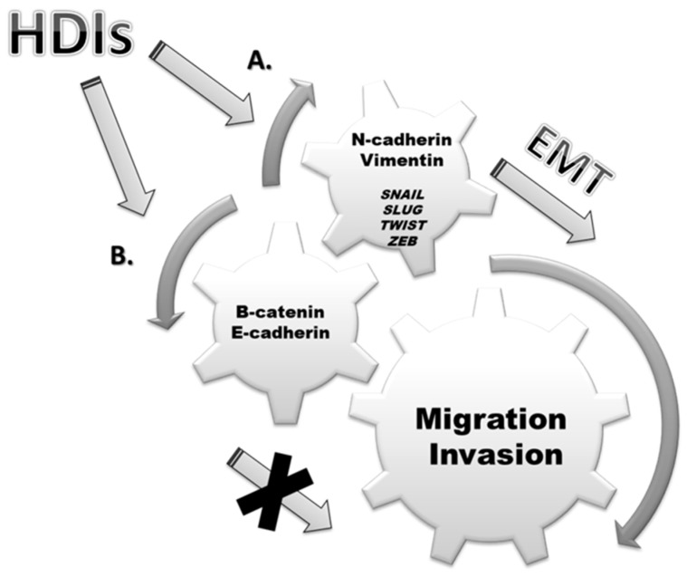
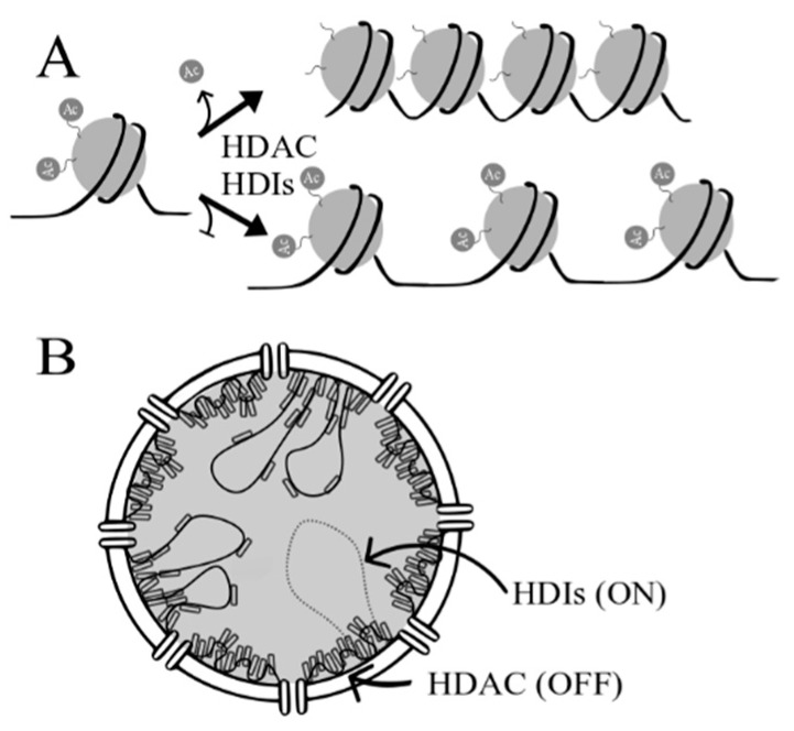
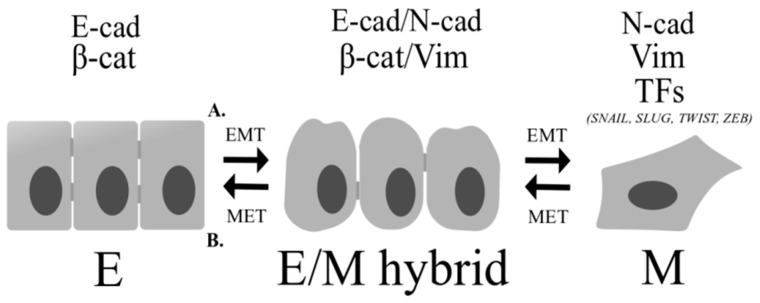
Histone deacetylase inhibitors (HDIs) are a group of potent epigenetic drugs which have been investigated for their therapeutic potential in various clinical disorders, including hematological malignancies and solid tumors. Currently, several HDIs are already in clinical use and many more are on clinical trials. HDIs have shown efficacy to inhibit initiation and progression of cancer cells. Nevertheless, both pro-invasive and anti-invasive activities of HDIs have been reported, questioning their impact in carcinogenesis. The aim of this review is to compile and discuss the most recent findings on the effect of HDIs on the epithelial-mesenchymal transition (EMT) process in human cancers. We have summarized the impact of HDIs on epithelial (E-cadherin, β-catenin) and mesenchymal (N-cadherin, vimentin) markers, EMT activators (TWIST, SNAIL, SLUG, SMAD, ZEB), as well as morphology, migration and invasion potential of cancer cells. We further discuss the use of HDIs as monotherapy or in combination with existing or novel anti-neoplastic drugs in relation to changes in EMT.

Keywords: EMT; HDAC; HDI; MET; cadherin; cancer; catenin; invasion; migration; vimentin.


Conflict of interest statement

The authors declare no conflict of interest.

Figures

Figure 1
Figure 1
Histone deacetylase inhibitors (HDIs) modulate expression of epithelial-mesenchymal transition (EMT) markers as well as stimulate or inhibit migration and invasion of cancer cells. (A) HDIs induce EMT by increasing migration and invasion of cancer cells by upregulation of mesenchymal markers (N-cadherin, vimentin) and EMT-related transcription factors (SNAIL, SLUG, TWIST, ZEB). (B) HDIs upregulate expression of epithelial markers (E-cadherin, β-catenin) and consequently inhibit EMT, migration and invasion of cancer cells.
Figure 2
Figure 2
(A) Effect of histone deacetylase inhibitors (HDIs) on chromatin remodeling. Acetylation (Ac) of histones results in changes in chromatin conformation, where non-acetylated histones form heterochromatin (close chromatin) while acetylated histones result in relaxed chromatin—allowing DNA-binding by transcription factors. (B) Chromosomal landscape in the nucleus in the presence and absence of HDIs. Closed chromatin is near the nuclear envelope, while relaxed chromatin, where transcription is possible, is found in the middle of nucleus.
Figure 3
Figure 3
Percentage (%) of patients with high or medium HDACs class I expression levels in different types of cancer [43,44,45,46].
Figure 4
Figure 4
Percentage (%) of patients with high or medium HDACs class II expression levels in different types of cancer. HDAC7 was not analyzed [47,48,49,50,51,52].
Figure 5
Figure 5
Percentage (%) of patients with high or medium HDACs class III expression levels in different types of cancer. There is no available data regarding the expression of HDAC11 (HDAC IV) [53,54,55,56,57,58,59,60].
Figure 6
Figure 6
Percentage (%) of tumors with high or medium HDAC protein expression levels in (A) breast cancer, (B) carcinoid, (C) cervical cancer, (D) colon cancer, (E) endometrial cancer [43,44,45,46,47,48,49,50,51,52,53,54,55,56,57,58,59,60].
Figure 7
Figure 7
Percentage (%) of tumors with high or medium HDAC protein expression levels in (A) glioma, (B) head and neck cancer, (C) liver cancer, (D) lung cancer, (E) lymphoma [43,44,45,46,47,48,49,50,51,52,53,54,55,56,57,58,59,60].
Figure 8
Figure 8
Percentage (%) of tumors with high or medium HDAC protein expression levels in (A) melanoma, (B) ovarian cancer, (C) pancreatic cancer, (D) prostate cancer, (E) renal cancer [43,44,45,46,47,48,49,50,51,52,53,54,55,56,57,58,59,60].
Figure 9
Figure 9
Percentage (%) of tumors with high or medium HDAC protein expression levels in (A) skin cancer, (B) stomach cancer, (C) testis cancer, (D) thyroid cancer, (E) urothelial cancer [43,44,45,46,47,48,49,50,51,52,53,54,55,56,57,58,59,60].
Figure 10
Figure 10
Phenotypical transformation of cells during the epithelial–mesenchymal transition (EMT) and mesenchymal-epithelial transition (MET) processes. (A) During EMT epithelial cells lose their polarized organization and acquire migratory and invasive capabilities by increase in mesenchymal markers (N-cadherin, vimentin) and EMT-related transcription factors (TFs) (SNAIL, SLUG, TWIST, ZEB). (B) During MET cells re-acquire epithelial properties. Epithelial cells are connected by intercellular junctions and they exhibit apical-basal polarization. The intermediate stage between fully-epithelial and fully-mesenchymal states has been described as E/M hybrid state. Cancer cells with E/M hybrid phenotype have cell-cell adhesion properties as well as migration abilities, simultaneously. E: epithelial; E/M hybrid: epithelial/mesenchymal hybrid; M: mesenchymal; E-cad: E-cadherin; β-cat: β-catenin; N-cad: N-cadherin; Vim: vimentin; TFs: transcription factors; SNAIL, SLUG, TWIST, ZEB: mesenchymal transcription factors.

References

    1. Davis F.M., Stewart T.A., Thompson E.W., Monteith G.R. Targeting EMT in cancer: Opportunities for pharmacological intervention. Trends Pharmacol. Sci. 2014;35:479–488. doi: 10.1016/j.tips.2014.06.006. - DOI - PubMed
    1. Kalluri R., Weinberg R.A. The basics of epithelial-mesenchymal transition. J. Clin. Investig. 2009;119:1420–1428. doi: 10.1172/JCI39104. - DOI - PMC - PubMed
    1. Fardi M., Solali S., Farshdousti Hagh M. Epigenetic mechanisms as a new approach in cancer treatment: An updated review. Genes Dis. 2018;5:304–311. doi: 10.1016/j.gendis.2018.06.003. - DOI - PMC - PubMed
    1. Ma F., Jiang S., Zhang C. Recent advances in histone modification and histone modifying enzyme assays. Expert Rev. Mol. Diagn. 2018:1–10. doi: 10.1080/14737159.2019.1559053. - DOI - PubMed
    1. Grabarska A., Łuszczki J.J., Nowosadzka E., Gumbarewicz E., Jeleniewicz W., Dmoszyńska-Graniczka M., Kowalczuk K., Kupisz K., Polberg K., Stepulak A. Histone Deacetylase Inhibitor SAHA as Potential Targeted Therapy Agent for Larynx Cancer Cells. J. Cancer. 2017;8:19–28. doi: 10.7150/jca.16655. - DOI - PMC - PubMed