Review
. 2019 Jan 27:70:91-104.
doi: 10.1146/annurev-med-121217-094234.

Structure-Based Vaccine Antigen Design

Affiliations
Review

Structure-Based Vaccine Antigen Design

Barney S Graham et al. Annu Rev Med. .

Abstract

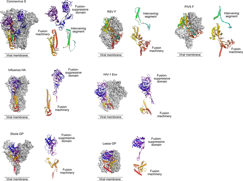
Enabled by new approaches for rapid identification and selection of human monoclonal antibodies, atomic-level structural information for viral surface proteins, and capacity for precision engineering of protein immunogens and self-assembling nanoparticles, a new era of antigen design and display options has evolved. While HIV-1 vaccine development has been a driving force behind these technologies and concepts, clinical proof-of-concept for structure-based vaccine design may first be achieved for respiratory syncytial virus (RSV), where conformation-dependent access to neutralization-sensitive epitopes on the fusion glycoprotein determines the capacity to induce potent neutralizing activity. Success with RSV has motivated structure-based stabilization of other class I viral fusion proteins for use as immunogens and demonstrated the importance of structural information for developing vaccines against other viral pathogens, particularly difficult targets that have resisted prior vaccine development efforts. Solving viral surface protein structures also supports rapid vaccine antigen design and application of platform manufacturing approaches for emerging pathogens.

Keywords: X-ray crystallography; coronavirus; electron microscopy; immunization; influenza; nanoparticle display; platform technology; respiratory syncytial virus; vaccine development.


Figures

Figure 1.
Figure 1.. Relative size and components of an antibody molecule.
(A) Full-length human IgG1 (PDB ID: 1IGT) is a dimer of heterodimers composed of two heavy chains (orange) and two light chains (light orange). One heavy and one light chain are shown in molecular surfaces and the other heavy and light chains are shown in ribbons. The variable (V) and constant (C) domains of both the light (L) and heavy (H) chains are labeled and the loops of the complementarity determining region (CDR) are highlighted in red and pink for the heavy and light chains, respectively. The antigen binding fragment (Fab) is distinguished from the crystallizable fragment (Fc). (B) The prefusion conformation of RSV F (pre-F) is shown with two protomers in grey and white molecular surfaces and a third protomer in ribbons, colored as a rainbow from blue to red, N-to C-terminus, respectively. Full-length IgG from (A) was aligned with the motavizumab Fab and is shown in molecular surfaces binding to pre-F, which is ~11 nm tall. The relative size of the IgG1 compared to the viral surface protein is demonstrated to illustrate how IgG binding to one epitope may partially interfere with binding to other nearby epitopes on the surface.
Figure 2.
Figure 2.. Epitopes present on two major conformations of the RSV F glycoprotein.
The prefusion conformation of RSV F (pre-F) is shown with two protomers in grey and white molecular surfaces and the third protomer in ribbons, colored as a rainbow from blue to red, N- to C- terminus, respectively. Antibodies against sites Ø (red/pink), III (green), and V (orange) bind only to pre-F, whereas antibodies that recognize sites II (yellow) and IV (purple) bind to regions that exist on the shared surfaces of pre-F and post-F. Antibodies to site I (blue) predominantly bind post-F. Antibodies that exclusively bind pre-F are the most potent, those that bind the shared surface tend to have moderate neutralizing activity, and those that bind only to post-F are typically non-neutralizing. Neutralization potency is illustrated by the width of the rainbow-colored triangle.
Figure 3.
Figure 3.. Class I viral fusion proteins.
Representative trimeric fusion proteins from selected virus families (Coronaviridae, Orthomyxoviridae, Filoviridae, Pneumoviridae, Retroviridae, Arenaviridae, and Paramyxoviridae) are shown with two protomers in grey and white molecular surfaces and a third protomer in ribbons, colored as described below. The panel adjacent to each trimer shows components related to the membrane fusion process, separated by function. These are type I membrane proteins anchored by a C-terminal transmembrane domain (TM). Each is shown in the pre-triggered state, with a free hydrophobic fusion peptide (FP) at the N-terminus of the C-terminal fragment, and two heptad-repeat regions adjacent to the FP and the TM that bring the viral and host-cell membrane together during the rearrangement process. These proteins vary in many ways, including whether there is a fusion-suppressive domain (colored from purple to blue) that must be displaced for the fusion machinery (colored from yellow to red) to function, and the presence or absence of an intervening segment that remains associated with the protein after fusion occurs (colored in cyan to green). Nevertheless, the overall functional similarities may lead to antigen design solutions that are analogous across viral families. Structures shown are derived from PDB IDs HCJ0, 2FK0, 5JQ3, 4MMU, 4ZMP, 5VK2, and 4GIP.

References

    1. Schmaljohn AL. 2013. Protective antiviral antibodies that lack neutralizing activity: precedents and evolution of concepts. Curr HIV Res 11: 345–53 - PubMed
    1. Ackerman ME, Alter G. 2013. Opportunities to exploit non-neutralizing HIV-specific antibody activity. Curr HIV Res 11: 365–77 - PMC - PubMed
    1. Sullivan NJ, Hensley L, Asiedu C, et al. 2011. CD8+ cellular immunity mediates rAd5 vaccine protection against Ebola virus infection of nonhuman primates. Nat Med 17: 1128–31 - PubMed
    1. McMichael AJ, Koff WC. 2014. Vaccines that stimulate T cell immunity to HIV-1: the next step. Nat Immunol 15: 319–22 - PMC - PubMed
    1. Kong L, Lee JH, Doores KJ, et al. 2013. Supersite of immune vulnerability on the glycosylated face of HIV-1 envelope glycoprotein gp120. Nat Struct Mol Biol 20: 796–803 - PMC - PubMed

Publication types

MeSH terms

LinkOut - more resources