. 2019 Jan 28;38(1):42.
doi: 10.1186/s13046-018-1008-8.

Activation of dynamin-related protein 1 - dependent mitochondria fragmentation and suppression of osteosarcoma by cryptotanshinone

Affiliations

Activation of dynamin-related protein 1 - dependent mitochondria fragmentation and suppression of osteosarcoma by cryptotanshinone

Jia-Hau Yen et al. J Exp Clin Cancer Res. .

Abstract

Background: Discovering how to regulate mitochondrial function to reduce cancer growth holds great potential for future cancer therapy development. Here we explore the effects of cryptotanshinone (CPT), a natural product derived from Salvia miltiorrhiza, on mitochondria of osteosarcoma (OS) both in vitro and in vivo, and further elucidate the underlying molecular mechanisms.

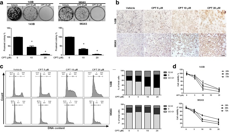
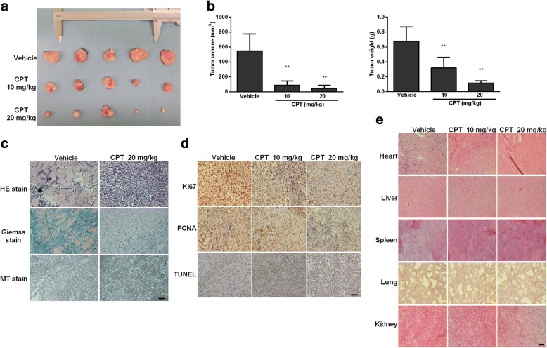
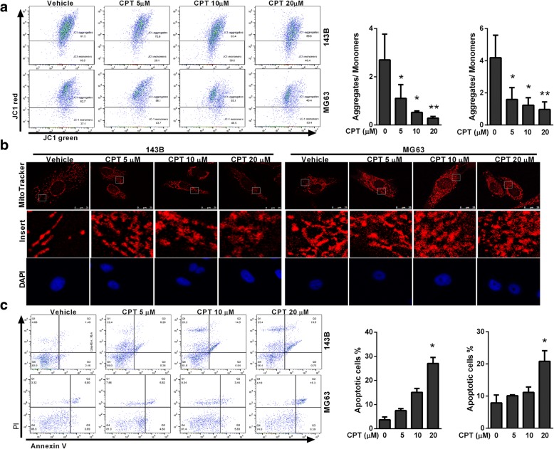
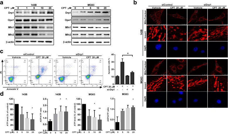
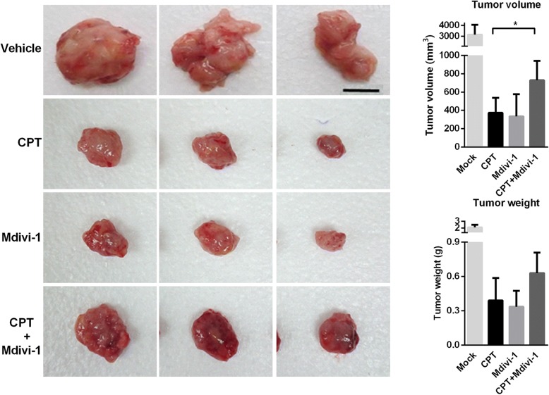
Methods: Cytotoxicity in the CPT treated OS cells was analyzed by flow cytometry, CCK8, TUNEL assay and colony formation assays. Flow cytometric analysis was performed to evaluate the effect of CPT on cell cycle of OS cells. Mitochondrial morphology was examined by staining with the mitochondrial membrane potential -sensitive fluorochrome, MitoTracker Red (CMXRos). Immunoblotting, confocal-immunofluorescence staining, co-immunoprecipitation were used to examine the expression and interaction between CPT-mediated Drp1 and Bax. Finally, the synergistic effect of CPT on OS cells was validated using a mouse xenograft tumor model.

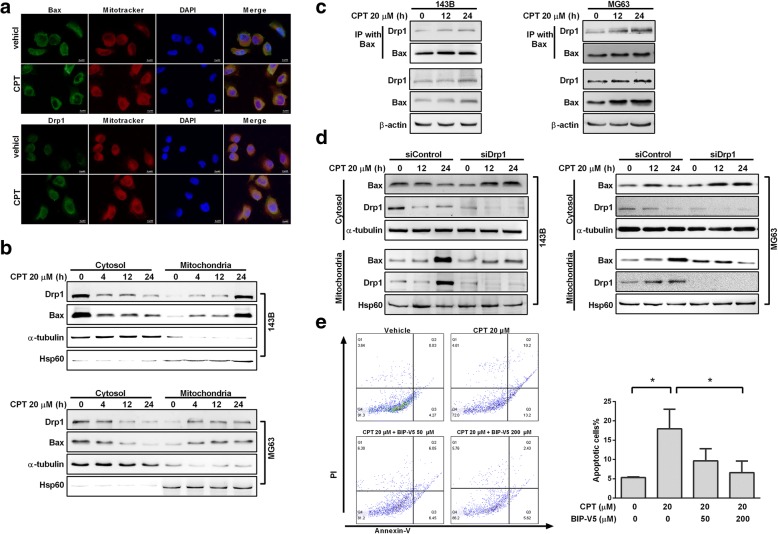
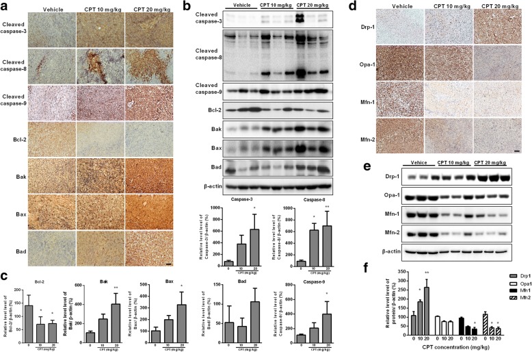
Results: In this study, we found CPT treatment induced S-phase arrest, apoptosis, and mitochondrial fragmentation in OS cells. CPT also effectively activated caspase-dependent apoptosis, which could be blocked by pan-caspase inhibitor Z-VAD-FMK. Moreover, we herein provide evidence that treatment with CPT resulted in mitochondrial fragmentation, which is mediated by dynamin-related protein 1 (Drp1), a key mediator of mitochondrial fission. Pursuing this observation, downregulation of Drp1 via silencing RNA could abrogate the induction of apoptosis and mitochondrial fragmentation induced by CPT. Finally, we demonstrate that CPT induced Drp1, which interacted directly with Bcl-2-associated X protein (Bax), which contributed to driving Bax translocation from the cytosol to the mitochondria.

Conclusions: Our findings offer insight into the crosstalk between mitochondrial fragmentation and inhibition of osteosarcoma cell growth in response to CPT.

Keywords: Cryptotanshinone (CPT); Drp1; Mitochondria fragmentation; Osteosarcoma.


Conflict of interest statement

Ethics approval and consent to participate

Not applicable.

Consent for publication

Not applicable.

Competing interests

The authors declare that they have no competing interests.

Publisher’s Note

Springer Nature remains neutral with regard to jurisdictional claims in published maps and institutional affiliations.

Figures

Fig. 1
Fig. 1
CPT induces S phase arrest and cells death in human OS cells. a Clonogenicity of OS cells treated with various concentrations of CPT (as indicated). b Representative images of TUNEL staining in OS cells treated with various concentrations of CPT (as indicated). Bar represents 50 μm. c OS cells were treated with control and CPT (as indicated) for 24 h. Flow-cytometric analysis and quantification of distribution of cell cycle were assessed. d OS cell viability following treatment with the various concentrations of CPT for 24, 48, and 72 h. CCK-8 assay was used to assess OS cell proliferation. The results were expressed as the means ± SD from three independent experiments. *P < 0.05, significantly different compared with control
Fig. 2
Fig. 2
In vivo evidence for CPT inhibits OS growth. a-b 143B cell-derived tumors were developed in nude mice and treated with vehicle or CPT. Tumor growth was monitored by measuring the tumor volume and tumor weight for 45 days (n = 5 mice/group; **P < 0.01). Representative tumor images are shown here. c Tumors dissected from vehicle and CPT-treated mice were subjected to H&E, Giemsa and Masson’s trichrome (MT) stains. dTumors were subjected to immunohistochemical analysis using Ki-67 and PCNA antibodies. Determination of apoptosis in tumor tissues by TUNEL assay. e Administration of CPT exhibited no toxicity in five major organs. H&E staining was used to evaluate the histology. Bar represents 500 μm
Fig. 3
Fig. 3
CPT triggers mitochondrial fragmentation and apoptosis in OS cells. a Representative dot plots of JC-1 aggregates versus JC-1 monomers in response to various CPT treatments of OS cells. b Representative changes in mitochondrial morphology were detected by confocal microscopy at various concentrations of CPT (as indicated). Images of mitochondria (red) and nucleus (blue) were collected by confocal microscope. c Annexin V/PI analysis by flow cytometry followed by CPT treatment (as indicated). The respective cell percentages in early and late apoptosis for different dose treatment are presented in the qantitative analysis. The results were expressed as the means ± SD from three independent experiments. *P < 0.05 and **P < 0.01, significantly different compared with control
Fig. 4
Fig. 4
Caspase-3, − 8, and − 9 are involvement in CPT-induced OS cell apoptosis. a The expressions of apoptosis-related proteins were measured by western blotting in OS cells following 20 μM CPT treatment for 24 h. b OS cells were pretreated with Z-VAD-FMK (20 μM) for 1 h and incubated with 20 μM CPT for 24 h. Annexin V/PI analysis was used to detect apoptotic cells by flow cytometry followed by CPT treatment. The respective cell percentages in early and late apoptosis for different dose treatment are presented in the quantitative analysis. The results were expressed as the means ± SD from three independent experiments. **P < 0.01, significantly different compared with control
Fig. 5
Fig. 5
Drp1 silencing attenuates CPT-induced mitochondrial fragmentation and apoptosis. a The effects of CPT on mitochondrial fusion/fission proteins expression in OS cells. Representative immunoblot of the protein levels of Drp1, Opa1, Mfn1 and Mfn2. b Comparison of mitochondrial morphology in the control and Drp1 siRNA cells. Images of mitochondria (red) and nucleus (blue) were collected by confocal microscope. c Drp1 silencing reversed the reduction of cell viability in CPT treated 143B cells. Representative dot plots of Annexin V/PI analysis after CPT treatments in the presence or absence of Drp1 siRNA. d CPT decreased ATP production and increased ADP/ATP ratio of both 143B and MG63 cells. The results were expressed as the means ± SD from three independent experiments. *P < 0.05, significantly different compared with control
Fig. 6
Fig. 6
Drp1 is required for CPT-mediated apoptosis. a Immuno-staining of Bax and Drp1. Confocal microscope scanning photographs indicated the mitochondrial localization of Drp1 and Bax in CPT treated 143B cells. Red: Mito-Tracker Red CMXRos, Green: Bax and Drp1 as indicated. b Expression of Drp1 and Bax in cytosolic and mitochondrial fractions in OS cells were determined by immunoblotting using anti-Drp1 and anti-Bax antibodies. c Interaction between Bax and Drp1 after CPT treatment. Proteins were extracted from CPT-treated OS cells, and then IP was performed with Bax antibody; Co-IP Drp1 and Bax was detected by Western blotting. d After knocking down Drp1 for 24 h, OS cells were treated with CPT 20 μM for indicated time. Bax expression was determined in both cytosolic and mitochondrial fractions using mouse anti-Bax antibody. Hsp60 and α-tubulin antibodies were used as loading controls for mitochondria and cytosol, respectively. e 143B cells were pretreated with Bax inhibitor peptide V5 (BIP-V5) (50 or 200 μM) for 1 h and incubated with 20 μM CPT for 24 h. Annexin V/PI analysis was used to detect apoptotic cells by flow cytometry followed by CPT treatment. The respective cell percentages in early and late apoptosis for different dose treatment are presented in the qantitative analysis. The results were expressed as the means ± SD from three independent experiments. *P < 0.05, significantly different compared with control
Fig. 7
Fig. 7
CPT activates caspase-mediated apoptosis and impaired balance of mitochondrial fission and fusion in vivo. 143B cell-derived tumors were developed in NOD/SCID mice and treated with CPT or vehicle for 45 days. Expressions of cleaved caspase 3, cleaved caspase 8, cleaved caspase 9, Bcl-2, Bak, Bax and Bad were examined by immunohistochemistry (a) and Western blotting (b). The quantification analysis in Figs. b is for Figs. (c). Expressions of Drp1, Opa1, Mfn1 and Mfn2 were examined by immunohistochemistry (d) and Western blotting (e). The quantification analysis in Figs. e is for Figs. (f). Ratios of each protein to β-actin were determined by densitometry. The results were expressed as the means ± SD from three independent experiments. *P < 0.05 and **P < 0.01, significantly different compared with control. Bar represents 500 μm.
Fig. 8
Fig. 8
Antitumor ability of CPT was Drp1 dependent. Efficacy of CPT in combination with Mdivi-1 in xenograft model bearing 143B cells. 143B cells were inoculated subcutaneously in NOD/SCID mice, tumors were allowed to form, and then mice were treated with CPT combined with or without Mdivi-1 (0.5 mg/mice). Representative tumor images are shown here. Tumor volumes and tumor weight were examined throughout the experiment. (n = 3 mice/group; *P < 0.05). Bar represents 1 cm

References

    1. Savage SA, Mirabello L. Using epidemiology and genomics to understand osteosarcoma etiology. Sarcoma. 2011;2011:548151. doi: 10.1155/2011/548151. - DOI - PMC - PubMed
    1. Chan DC. Mitochondria: dynamic organelles in disease, aging, and development. Cell. 2006;125(7):1241–1252. doi: 10.1016/j.cell.2006.06.010. - DOI - PubMed
    1. Rafelski SM. Mitochondrial network morphology: building an integrative, geometrical view. BMC Biol. 2013;11:71. doi: 10.1186/1741-7007-11-71. - DOI - PMC - PubMed
    1. Germain M, Mathai JP, McBride HM, Shore GC. Endoplasmic reticulum BIK initiates DRP1-regulated remodelling of mitochondrial cristae during apoptosis. EMBO J. 2005;24(8):1546–1556. doi: 10.1038/sj.emboj.7600592. - DOI - PMC - PubMed
    1. Chen H, Chomyn A, Chan DC. Disruption of fusion results in mitochondrial heterogeneity and dysfunction. J Biol Chem. 2005;280(28):26185–26192. doi: 10.1074/jbc.M503062200. - DOI - PubMed

MeSH terms