. 2019:616:61-85.
doi: 10.1016/bs.mie.2018.10.027. Epub 2018 Dec 21.

Fluorescence-based methods for measuring target interference by CRISPR-Cas systems

Affiliations

Fluorescence-based methods for measuring target interference by CRISPR-Cas systems

Phong T Phan et al. Methods Enzymol. 2019.

Abstract

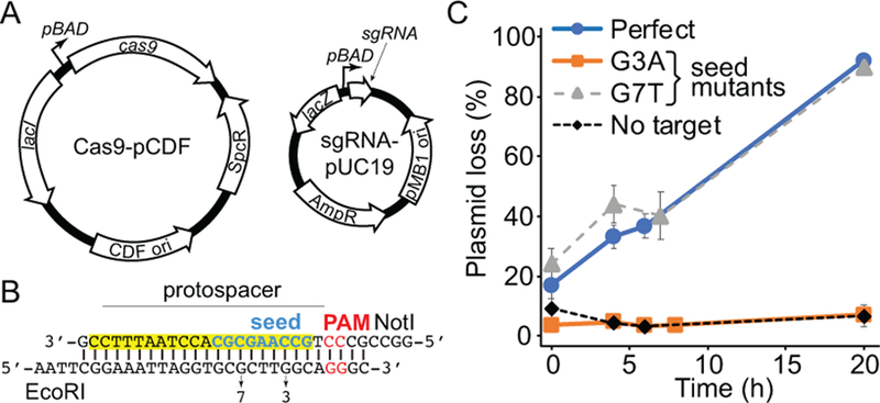
Type I, II, and V CRISPR-Cas systems are RNA-guided dsDNA targeting defense mechanisms found in bacteria and archaea. During CRISPR interference, Cas effectors use CRISPR-derived RNAs (crRNAs) as guides to bind complementary sequences in foreign dsDNA, leading to the cleavage and destruction of the DNA target. Mutations within the target or in the protospacer adjacent motif can reduce the level of CRISPR interference, although the level of defect is dependent on the type and position of the mutation, as well as the guide sequence of the crRNA. Given the importance of Cas effectors in host defense and for biotechnology tools, there has been considerable interest in developing sensitive methods for detecting Cas effector activity through CRISPR interference. In this chapter, we describe an in vivo fluorescence-based method for monitoring plasmid interference in Escherichia coli. This approach uses a green fluorescent protein reporter to monitor varying plasmid levels within bacterial colonies, or to measure the rate of plasmid-loss in bacterial populations over time. We demonstrate the use of this simple plasmid-loss assay for both chromosomally integrated and plasmid-borne CRISPR-Cas systems.

Keywords: CRISPR interference; CRISPR–Cas; Cas9; Cascade; Flow cytometry; Fluorescence imaging; GFP; Molecular biology; Typhoon imager.


Figures

Figure 1
Figure 1. Design of fluorescence-based CRISPR interference assay.
A. Schematic of Cas-crRNA effector complex bound to dsDNA. The spacer-protospacer RNA:DNA hybrid is shown in yellow. The PAM is highlighted in red. The PAM-proximal seed region is labeled. B. Schematic of pACYC-GFP plasmid. MCS1: multiple cloning site 1. C. Close-up schematic of GFP expression cassette in pACYC-GFP. The locations of the constitutive promoter and ssrA degradation tag are highlighted. D. Basis for fluorescence-based plasmid loss assay. As the plasmid concentration decreases due to CRISPR-based plasmid loss, the cells become less fluorescent.
Figure 2
Figure 2. Development of pACYC-GFP.
A. Constitutive promoter sequences tested during construction of pACYC-GFP. Promoters 1–4 are variants of tac promoter that reduce the promoter strength. Promoter 1 contains an extra base pair between the −10 and −35 sites. Promoters 2, 3 and 4 contain one, two or three variations in the −35 site, respectively. Variations between promoters are underlined. Gaps in aligned promoter sequences are represented with spaces. B. Flow cytometry histograms for the five promoters tested. p1-p4: promoters 1–4. C. Competition assays between cells harboring pACYCDuet-1 (GFP−) and pACYC-GFP (GFP+). The population distribution remains the same after 24 h of growth, indicating that pACYC-GFP does not affect growth rate of the cells.
Figure 3
Figure 3. Detecting CRISPR interference in bacterial colonies.
A. Design of target sequence inserted into pACYC-GFP. The perfect target is shown, similar oligonucleotides bearing G1C, A4G, AAA PAM or AGA PAM (non-target strand sequences) mutations were used for mutant target sequences. Positions of seed mutations are indicated. The target-strand protospacer is highlighted in yellow, the seed in blue, and the PAM in red. NcoI and NotI overhangs are labeled. B. Typhoon scanned plates for perfect target, empty pACYC-GFP lacking a CRISPR target, and the four mutant target plasmids. C. Box plot of quantified intensities for colonies on each plate. The mean intensity for each colony was normalized against the average mean intensity for colonies from the empty pACYC-GFP plate ([mean intensity induced colony]/[average mean intensity for all empty pACYC-GFP colonies]). Boxes depict variation from 25th to 75th percentile with the line within the box representing the median value and the X marking the mean. Error bars depict the local minimum and maximum, outliers are shown as circles.
Figure 4
Figure 4. Monitoring CRISPR interference over time in liquid cultures.
Plasmid loss (%) is the percentage of GFP- cells based on flow cytometry measurements at each time point. The average plasmid loss from 2–4 replicates is shown, with error representing standard deviation.
Figure 5
Figure 5. Measurement of Cas9 cleavage using GFP reporter assay.
A. Schematic of cas9 and gRNA expression plasmids. The cas9 gene and sgRNA are both expressed from arabinose inducible pBAD promoters. B. Target inserted into pACYC-GFP for this study. The target-strand protospacer is highlighted in yellow, the seed in blue, and the PAM in red. EcoRI and NotI overhangs are labeled. Positions of G3A or G7T (non-target strand sequence) mutations are indicated. C. Plasmid loss assay for Cas9 targets containing a perfect sequence or seed mismatches at the third or seventh position. Empty pACYC-GFP (no target) was used as a control to ensure that the plasmid is stable in the absence of CRISPR interference. Plasmid loss (%) is the percentage of GFP- cells based on flow cytometry measurements at each time point. The average plasmid loss from 3 replicates is shown, with error representing standard deviation.

References

    1. Abudayyeh OO, Gootenberg JS, Konermann S, Joung J, Slaymaker IM, Cox DBT, … Zhang F (2016). C2c2 is a single-component programmable RNA-guided RNA-targeting CRISPR effector. Science, 353(6299). 10.1126/science.aaf5573 - DOI - PMC - PubMed
    1. Andersen JB, Sternberg C, Poulsen LK, Bjørn SP, Givskov M, & Molin S (1998). New Unstable Variants of Green Fluorescent Protein for Studies of Transient Gene Expression in Bacteria. Applied and Environmental Microbiology, 64(6), 2240 LP-2246. - PMC - PubMed
    1. Brouns SJJ, Jore MM, Lundgren M, Westra ER, Slijkhuis RJH, Snijders APL, … van der Oost J (2008). Small CRISPR RNAs guide antiviral defense in prokaryotes. Science, 321(5891), 960–4. 10.1126/science.1159689 - DOI - PMC - PubMed
    1. Cooper LA, Stringer AM, & Wade JT (2018). Determining the Specificity of Cascade Binding, Interference, and Primed Adaptation In Vivo in the Escherichia coli Type I-E CRISPR-Cas System. MBio 10.1101/169011 - DOI - PMC - PubMed
    1. Datsenko KA, Pougach K, Tikhonov A, Wanner BL, Severinov K, & Semenova E (2012). Molecular memory of prior infections activates the CRISPR/Cas adaptive bacterial immunity system. Nature Communications, 3, 945 10.1038/ncomms1937 - DOI - PubMed

Publication types

MeSH terms

Substances

LinkOut - more resources