. 2019 Feb 12;3(3):242-255.
doi: 10.1182/bloodadvances.2018024182.

Selective targeting of NAMPT by KPT-9274 in acute myeloid leukemia

Affiliations

Selective targeting of NAMPT by KPT-9274 in acute myeloid leukemia

Shaneice R Mitchell et al. Blood Adv. .

Abstract

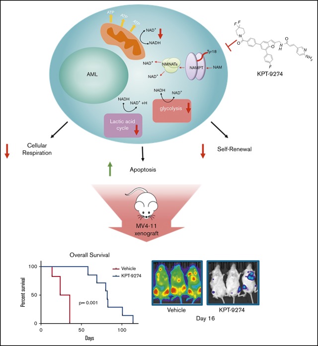
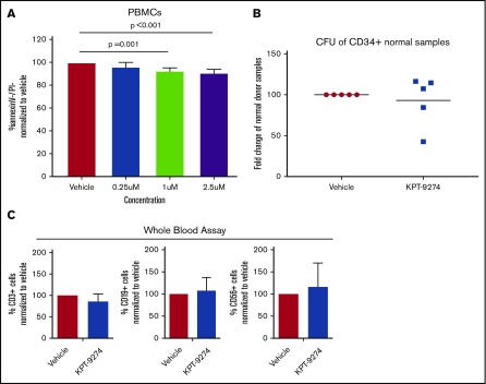
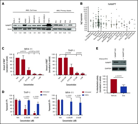
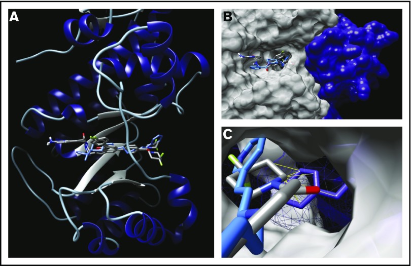
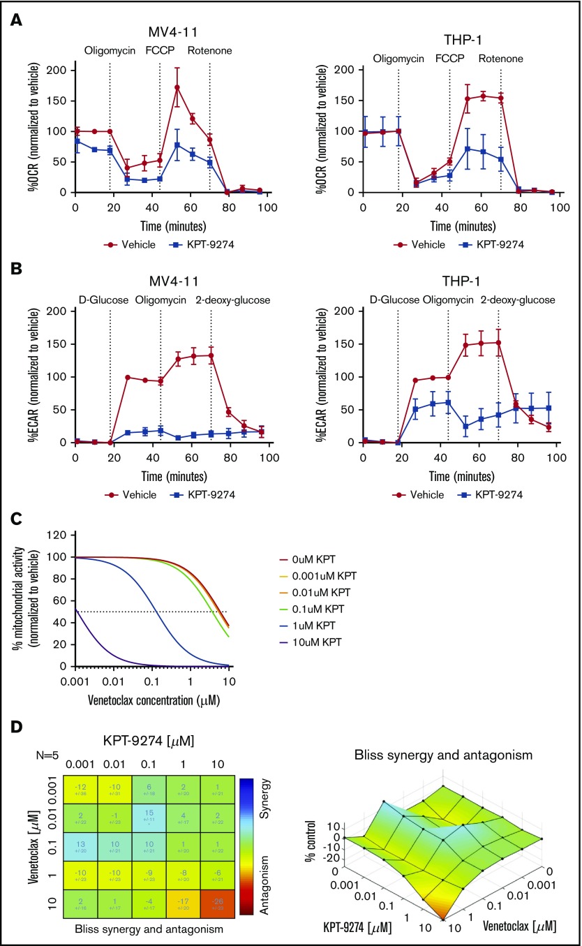
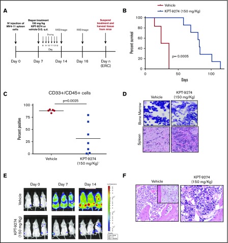
Treatment options for acute myeloid leukemia (AML) remain extremely limited and associated with significant toxicity. Nicotinamide phosphoribosyltransferase (NAMPT) is involved in the generation of NAD+ and a potential therapeutic target in AML. We evaluated the effect of KPT-9274, a p21-activated kinase 4/NAMPT inhibitor that possesses a unique NAMPT-binding profile based on in silico modeling compared with earlier compounds pursued against this target. KPT-9274 elicited loss of mitochondrial respiration and glycolysis and induced apoptosis in AML subtypes independent of mutations and genomic abnormalities. These actions occurred mainly through the depletion of NAD+, whereas genetic knockdown of p21-activated kinase 4 did not induce cytotoxicity in AML cell lines or influence the cytotoxic effect of KPT-9274. KPT-9274 exposure reduced colony formation, increased blast differentiation, and diminished the frequency of leukemia-initiating cells from primary AML samples; KPT-9274 was minimally cytotoxic toward normal hematopoietic or immune cells. In addition, KPT-9274 improved overall survival in vivo in 2 different mouse models of AML and reduced tumor development in a patient-derived xenograft model of AML. Overall, KPT-9274 exhibited broad preclinical activity across a variety of AML subtypes and warrants further investigation as a potential therapeutic agent for AML.


Conflict of interest statement

Conflict-of-interest disclosures: E.B. and W.S. are employees of, and have financial interests in, Karyopharm Therapeutics Inc. J.S.B. has performed consulting for AbbVie, AstraZeneca, and Kite Pharma. V.K.P. performed consulting for Orbus Therapeutics and SK Biosciences. The remaining authors declare no competing financial interests.

Figures

None
Graphical abstract
Figure 1.
Figure 1.
KPT-9274 significantly inhibits proliferation and induces apoptosis of AML cell lines. (A) Apoptosis as measured by using annexin V/propidium iodide (PI) flow cytometric analysis at 24, 48, and 72 hours in 5 AML cell lines treated with KPT-9274. (B) Caspase-9, caspase-3, and poly (ADP-ribose) polymerase (PARP) cleavage after treatment of KPT-9274 at 24, 48, and 72 hours in MV4-11 and THP-1 cell lines. (C) Colony assays using patient primary cells (n = 11) after KPT-9274 treatment and 14 days of plating. (D) Replating of patient primary samples (n = 6) after treatment and 14 days of plating in MethoCult medium.
Figure 2.
Figure 2.
KPT-9274 spares normal hematopoietic cells from toxicity, suggesting selective toxicity. (A) Annexin V/propidium iodide (PI) analysis of 5 normal donor peripheral blood mononuclear cells (PMBCs) treated with KPT-9274 after 72 hours. (B) Treatment with 250 nM of KPT-9274 revealed no significant decrease in colony formation of normal donor CD34+ stem cells (n = 4). (C) Whole-blood samples from normal donors (n = 5) were treated with or without 1 µM of KPT-9274 and accessed for viability of normal lymphocytes after 48 hours of treatment using flow cytometric analysis.
Figure 3.
Figure 3.
NAMPT as the primary target of KPT-9274 in AML. (A) Variable expression of NAMPT protein in AML cell lines and patient samples. (B) Expression levels in AML patient samples with common genetic aberrations (Bloodspot; The Cancer Genome Atlas AML data). (C) MV4-11 and THP-1 cells treated with KPT-9274 were assessed for NAD+ reduction after 24 hours of treatment (n = 3). (D) Annexin V/propidium iodide (PI) flow cytometric analysis of KPT-9274–treated cell lines MV4-11 and THP-1 after addition of 100 µM of exogenous NAD+. (E) Western blot analysis showing the degree of CRISPR-Cas9 knockdown (KD) of NAMPT in THP-1 cells after 120 hours of doxycycline (dox) treatment. Graph shows mitochondrial activity analysis of cell lines treated with doxycycline of CRISPR-Cas9 NAMPT knockdown THP-1 cell line using MTS (n = 3).
Figure 4.
Figure 4.
Docking of KPT-9274 into NAMPT. (A) Two lowest energy docked conformers of KPT-9274 (gray) overlaid on FK866 (blue) showing consistent binding mode with alternate binding modes for the piperidine end of KPT-9274. (B) NAMPT homodimer shown using surface representation, chain A (white) depicted at binding cavity with KPT-9274 (gray) and FK866 (blue) complexed to chain B (purple). (C) Detailed view of distal tunnel containing bound inhibitors showing favorable ring-stacking interaction between FK866 and tyrosine residue of chain B compared with the steric clash of KPT-9274 with the same tyrosine.
Figure 5.
Figure 5.
NAMPT inhibition impairs cellular respiration in AML cells. (A) Oxygen consumption rate (OCR) measured before and after the addition of the inhibitors oligomycin, carbonyl cyanide 4-(trifluoromethoxy)phenylhydrazone (FCCP), and rotenone to derive several parameters of mitochondrial respiration following KPT-9274 treatment (24 hours, 250 nM; n = 3). (B) Results of glycolytic stress testing using MV4-11 and THP-1 cell lines (24 hours, 250 nM; n = 3). (C) Mitochondrial activity of patient samples treated with venetoclax given in combination with different doses of KPT-9274 in the presence of stromal protection (n = 5; error bars were removed for clarity). (D) Analysis of synergistic effect of combination of KPT-9274 with venetoclax, showing significant synergy at the 100-nM dose of KPT-9274 in combination with the 10-nM dose of venetoclax using Combenefit software analysis. ECAR, extracellular acidification rate.
Figure 6.
Figure 6.
KPT-9274 increases survival rates and prevents disease migration in an AML xenograft mouse model. (A) Dosing schematic of NSG mice engrafted with MV4-11 luciferase-expressing cells were treated with 150 mg/kg of KPT-9274 (n = 7) or vehicle control (n = 5). (B) Overall survival of KPT-9274–treated mice vs vehicle-treated mice are shown according to Kaplan-Meier analysis. (C) Human CD33+/CD45+ cells were assessed in the bone marrow at the end of the study by using flow cytometry. (D) Cytology of bone marrow cytocentrifugation preparations (top row; 100× oil objective, Wright-Giemsa stain) and histology of the spleen (bottom row; 60× magnification, hematoxylin and eosin stain) of KPT-9274–treated and vehicle-treated mice who met euthanasia removal criteria and were removed from the study. (E) Disease progression was assessed in a separate cohort of mice treated with KPT-9274 (n = 3) and vehicle control (n = 3) by using IVIS bioluminescence after treatment began. (F) Histopathology of the bone marrow of mice euthanized before mice met early removal criteria (ERC) in KPT-9274–treated and vehicle-treated mice (60× magnification, hematoxylin and eosin stain). Bone marrow from vehicle-treated mice revealed multifocal infiltration by neoplastic myeloid cells, with occasional foci of coagulation necrosis or infarction, presumably due to outgrowth of blood supply (inset). By contrast, mice treated with KPT-9274 showed markedly less or absent infiltration. O.G., oral gavage; q.d., once daily.
Figure 7.
Figure 7.
KPT-9274 decreases disease burden and infiltration in a PDX model of AML. (A) Mice with confirmed engraftment were treated for 4 weeks with 150 mg/kg of KPT-9274 oral gavage, once daily (n = 7) or vehicle control (n = 8). (B) At the end of the study, human CD33+ cells were assessed by using flow cytometry in the bone marrow in both groups. (C) Whole blood analysis of human CD33+ cells in mice after 4 weeks. (D-E) Spleen weights of KPT-9274–treated (n = 7) and vehicle-treated (n = 6) mice. (F) Histology images from PDX mice after 4 weeks of treatment with KPT-9274 or vehicle control. Mice treated with KPT-9274 showed the least severe infiltration of liver, spleen, and bone marrow, whereas infiltration of the liver was often absent in this group (liver, spleen, and bone marrow: 40× magnification, hematoxylin and eosin stain). Vehicle-treated mice had the most severe infiltration of the spleen, bone marrow, and liver. Differentials were performed on cytospins of bone marrow aspirates, which showed myeloid differentiation and fewer blasts in KPT-9274–treated mice vs the vehicle control group (100× oil objective, Wright-Giemsa stain). Leukemia cells are indicated with asterisks (*). (G) Assessment of CD34+/CD38 fraction of leukemic cells in vehicle-treated and KPT-9274–treated mice. (H) Colony formation assays of KPT-9274–treated mouse samples vs vehicle control group after primary, secondary, and tertiary replating. BM, bone marrow.

References

    1. Estey E, Döhner H. Acute myeloid leukaemia. Lancet. 2006;368(9550):1894-1907. - PubMed
    1. Döhner H, Estey EH, Amadori S, et al. ; European LeukemiaNet. Diagnosis and management of acute myeloid leukemia in adults: recommendations from an international expert panel, on behalf of the European LeukemiaNet. Blood. 2010;115(3):453-474. - PubMed
    1. Appelbaum FR, Gundacker H, Head DR, et al. . Age and acute myeloid leukemia. Blood. 2006;107(9):3481-3485. - PMC - PubMed
    1. Levis M. Midostaurin approved for FLT3-mutated AML. Blood. 2017;129(26):3403-3406. - PubMed
    1. Stone RM, Mandrekar SJ, Sanford BL, et al. Midostaurin plus chemotherapy for acute myeloid leukemia with a FLT3 mutation. N Engl J Med 2017;377(5):454-464. - PMC - PubMed

Publication types