. 2019 Mar 26;201(8):e00698-18.
doi: 10.1128/JB.00698-18. Print 2019 Apr 15.

Roles of the DedD Protein in Escherichia coli Cell Constriction

Affiliations

Roles of the DedD Protein in Escherichia coli Cell Constriction

Bing Liu et al. J Bacteriol. .

Abstract

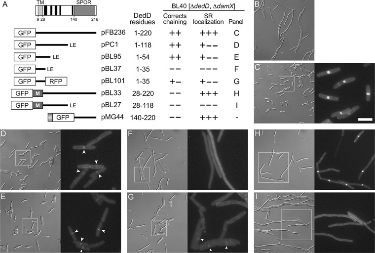
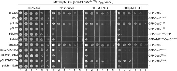
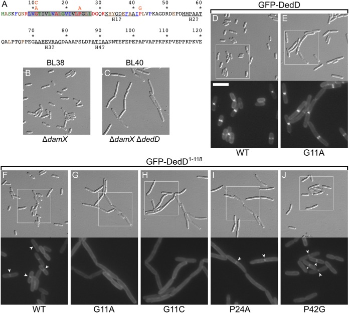
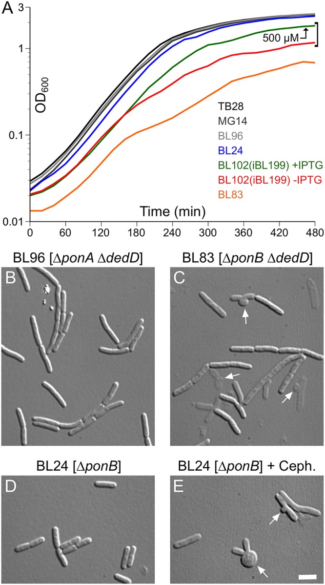
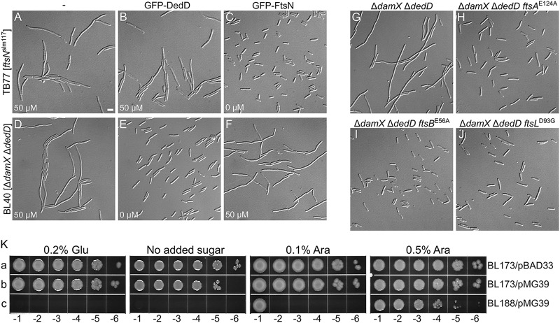
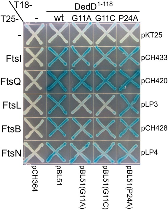
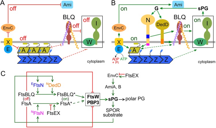
Two key tasks of the bacterial septal-ring (SR) machinery during cell constriction are the generation of an inward-growing annulus of septal peptidoglycan (sPG) and the concomitant splitting of its outer edge into two layers of polar PG that will be inherited by the two new cell ends. FtsN is an essential SR protein that helps trigger the active constriction phase in Escherichia coli by inducing a self-enhancing cycle of processes that includes both sPG synthesis and splitting and that we refer to as the sPG loop. DedD is an SR protein that resembles FtsN in several ways. Both are bitopic inner membrane proteins with small N-terminal cytoplasmic parts and larger periplasmic parts that terminate with a SPOR domain. Though absence of DedD normally causes a mild cell-chaining phenotype, the protein is essential for division and survival of cells with limited FtsN activity. Here, we find that a small N-terminal portion of DedD (NDedD; DedD1-54) is required and sufficient to suppress ΔdedD-associated division phenotypes, and we identify residues within its transmembrane domain that are particularly critical to DedD function. Further analyses indicate that DedD and FtsN act in parallel to promote sPG synthesis, possibly by engaging different parts of the FtsBLQ subcomplex to induce a conformation that permits and/or stimulates the activity of sPG synthase complexes composed of FtsW, FtsI (PBP3), and associated proteins. We propose that, like FtsN, DedD promotes cell fission by stimulating sPG synthesis, as well as by providing positive feedback to the sPG loop.IMPORTANCE Cell division (cytokinesis) is a fundamental biological process that is incompletely understood for any organism. Division of bacterial cells relies on a ring-like machinery called the septal ring or divisome that assembles along the circumference of the mother cell at the site where constriction eventually occurs. In the well-studied bacterium Escherichia coli, this machinery contains over 30 distinct proteins. We identify functionally important parts of one of these proteins, DedD, and present evidence supporting a role for DedD in helping to induce and/or sustain a self-enhancing cycle of processes that are executed by fellow septal-ring proteins and that drive the active constriction phase of the cell division cycle.

Keywords: FtsA; FtsB; FtsL; FtsQ; cell division.


Figures

FIG 1
FIG 1
Domain analyses of E. coli DedD. (A) Schematic depiction of full-length DedD (DedD1–220). The transmembrane domain (TMDedD) and the C-terminal SPOR domain (SDedD) are indicated. Potential α-helices (H1 to H5), predicted to form in the periplasmic portion between TMDedD and SDedD by the GOR IV secondary-structure prediction method (85), are indicated in black. Also shown are inserts present on plasmids that encode fusions of various portions of DedD to GFP or TTGFP under control of the lac regulatory region. The TTGFP fusion encoded by pMG44 contains the TorA signal peptide (hatched box) that is cleaved upon export to the periplasm via the twin arginine transport (Tat) system. M represents residues 2 to 39 of MalF, which include cytoplasmic residues and the first transmembrane domain (TM1; MalF19–35) of the protein (88). Some fusions end with a nonnative Leu-Glu dipeptide (LE), as indicated. The table on the right shows (left to right) the DedD residues present in each fusion; whether the fusion could fully (++) or partially (+−) or could not (−−) correct the cell-chaining phenotype of BL40 [ΔdamX ΔdedD]; whether the fusion accumulated sharply (+++), weakly (+−−), or not at all (−−−) at division sites in the strain; and the panel with corresponding cell images. (B to I) Images of live BL40 [ΔdamX ΔdedD] cells carrying the vector control pMLB1113ΔH [Plac::] (B), pFB236 [Plac::gfp-dedD] (C), pPC1 [Plac::gfp-dedD1–118] (D), pBL95 [Plac::gfp-dedD1–54] (E), pBL37 [Plac::gfp-dedD1–35] (F), pBL101 [Plac::gfp-dedD1–35-rfp] (G), pBL33 [Plac::gfp-malF2–39-dedD28–220] (H), or pBL27 [Plac::gfp-malF2–39-dedD28–118] (I). Panels C to I comprise a differential interference contrast (DIC) image (left) and a fluorescence (FL) image (right) that corresponds to the boxed area in the DIC image. Bar, 16 μm (all DIC images), 8 μm (FL images in panels H and I), or 4 μm (FL images in panels C to G). Cells were grown for ∼5 mass doublings to an OD600 of 0.5 to 0.6 in LB with 50 μM IPTG. Strong septal localization of TTGFP-DedD140–220 was shown previously (71) and resembled that seen in panel H. Examples of weak (GFP-DedD1–118 and GFP-DedD1–54) or very weak (GFP-DedD1–35-RFP) septal accumulations of some of the SPOR-less fusions are marked by arrowheads in panels D, E, and G.
FIG 2
FIG 2
Ability of GFP-DedD and variants to rescue DedD-depleted ftsNslm117 cells. Strain MG19/pMG39 [ΔdedD ftsNslm117/PBAD::dedD] carrying one of the indicated plasmids was grown overnight in LB with 0.5% arabinose. Cultures were serially diluted in LB to an OD600 of 4 × 10x, and 5 μl of each dilution was spotted on LB agar containing arabinose, IPTG, or neither, as indicated. The plates were incubated for 16 h. Save for the vector control, pMLB1113ΔH [Plac::], each plasmid encoded GFP-DedD or a mutant derivative under control of the lac regulatory region. The relevant plasmid name and encoded protein are indicated on the left and right of each row, respectively. Plasmids pFB236 and pBL272 are almost identical, except that the GFP-DedD fusion encoded by the latter carries an additional four residues in the linker peptide that connects the GFP and DedD moieties.
FIG 3
FIG 3
Phenotype of SPOR-less DedD. (A to J) DIC images of live cells of strains BL40(iBL360) [ΔdamX ΔdedD(Plac::gfp-dedD)] (A to E) and BL40(iBL345) [ΔdamX ΔdedD(Plac::gfp-dedD1–118)] (F to J). The cells were grown for ∼3 mass doublings to an OD600 of 0.5 to 0.6 in M9-maltose with 0 μM (A and F), 10 μM (B and G), 50 μM (C and H), 100 μM (D and I), or 250 μM (E and J) IPTG. Bar, 4 μm. (K) Western analysis of full-length GFP-DedD and SPOR-less GFP-DedD1–118. Nonchaining cells of strains BL38 [ΔdamX] (lane 1), BL40(iBL360) [ΔdamX ΔdedD(Plac::gfp-dedD)] (lane 2), and BL40(iBL345) [ΔdamX ΔdedD] [Plac::gfp-dedD1–118] (lane 3) were obtained by growth as described for panel C [50 μM IPTG; BL38 and BL40(iBL360)] or J [250 μM IPTG; BL40(iBL345)] and used to prepare whole-cell extracts. For lanes 4 to 7, the BL40(iBL345) extract (lane 3) was diluted with that of BL38 (lane 1) to yield the fraction of the former indicated above each lane. Each lane contained 40 μg total protein. Fusion proteins were detected using anti-GFP (α-GFP) polyclonal antibodies. Bands corresponding to the fusions of interest are indicated by arrowheads. Migration of molecular mass standards (in kilodaltons) is indicated on the left. Division phenotypes (A to J) and relative band intensities (K) indicated that, relative to GFP-DedD, BL40 [ΔdamX ΔdedD] cells require a 2- to 4-fold higher level of SPOR-less GFP-DedD1–118 to prevent cell chaining.
FIG 4
FIG 4
Identification of residues important for DedD function. (A) DedD residues 1 to 120 are colored according to phylogenetic conservation (percent identity in the Hogenom gene family HOG000279501 [90]); red, 100 to 98%; blue, 98 to 95%; green, 95 to 90%; brown, 90 to 75%; and black, <75%. DedD is a type II bitopic (N-in) inner membrane protein, and its transmembrane domain is shaded in gray. Pertinent substitutions (orange) are indicated immediately above the affected residues. (B and C) DIC images of live BL38 [ΔdamX] (B) and BL40 [ΔdamX ΔdedD] (C) cells after growth for ∼3 mass doublings to an OD600 of 0.5 to 0.6 in M9-maltose. (D to J) Images of live BL40 [ΔdamX ΔdedD] cells carrying a single copy of iBL360 [Plac::gfp-dedD] (D), iBL360(G11A) [Plac::gfp-dedDG11A] (E), iBL345 [Plac::gfp-dedD1–118] (F), iBL345(G11A) [Plac::gfp-dedD1–118, G11A] (G), iBL345(G11C) [Plac::gfp-dedD1–118, G11C] (H), iBL345(P24A) [Plac::gfp-dedD1–118, P24A] (I), or iBL345(P42G) [Plac::gfp-dedD1–118, P42G] (J) integrated in the chromosome. The cells were grown as for panels B and C, but with 250 μM IPTG included in the medium. The panels comprise a DIC image (top) and an FL image (bottom) that corresponds to the boxed area in the DIC image. Bar, 8 μm (DIC images) or 4 μm (FL images). The arrowheads in panels F, I, and J mark examples of the weak accumulation of GFP-DedD1–118 or mutant derivatives at division sites, which are not seen in panels G and H.
FIG 5
FIG 5
Absence of both DedD and PBP1B causes severe cell lysis. (A) Growth curves of strains TB28 [wt] (black), MG14 [ΔdedD] (dark gray), BL96 [ΔponA ΔdedD] (light gray), BL24 [ΔponB] (blue), BL83 [ΔponB ΔdedD] (orange), and the DedD depletion strain BL102(iBL199) [ΔponB ΔdedD(Plac::gfp-dedD1–54)] (red and green). Cultures were grown overnight in LB with 500 μM IPTG [BL102(iBL199)], or without inducer (all other strains) and diluted 200-fold in LB with 500 μM IPTG (green) or without inducer (all other curves). Growth was continued, and OD600 values were determined every 20 min. Note that the TB28, MG14, BL96, and BL24 curves almost coincide. The shape of the orange curve reflects the relatively low densities attained by overnight cultures of strain BL83 [ΔponB ΔdedD], a relatively long lag period, and a decrease in the rate of optical density increase around 160 min. (B to E) Strains BL96 [ΔponA ΔdedD] (B), BL83 [ΔponB ΔdedD] (C), and BL24 [ΔponB] (D and E) were cultured in parallel as in panel A, and the cells were imaged 3 h (B and D) (OD600, ∼0.6) or 5 h (C) (OD600, ∼0.3) after inoculation. When the culture in panel E reached an OD600 of 0.3, cephalexin was added to 15 μg/ml, and the cells were imaged 30 min later. The arrows in panels C and E indicate examples of septal bulges and rabbit ears, indicative of septal lysis. Bar, 4 μm.
FIG 6
FIG 6
DedD functions independently of FtsN. (A to J) Cells were grown for ∼5 mass doublings to an OD600 of 0.5 to 0.6 in LB with 50 μM IPTG (A, B, D, and F) or without inducer (C, E, and G to J) and were imaged live (A to F) or after chemical fixation (G to J). Bar, 4 μm. (A to F) Little to no cross-functionality of FtsN and DedD. Cells of strain TB77 [ftsNslm117] (A to C) and BL40 [ΔdamX ΔdedD] (D to F) carrying pMLB1113ΔH [Plac::] (A and D), pFB236 [Plac::gfp-dedD] (B and E), or pCH201 [Plac::gfp-ftsN] (C and F) are shown. (G to J) EFtsN*-suppressing mutations in ftsA, ftsB, or ftsL also suppress the division defect associated with the absence of DedD. Shown are cells of strains BL40 [ΔdamX ΔdedD] (G), CH178 [ΔdamX ΔdedD ftsAE124A] (H), CH177 [ΔdamX ΔdedD ftsBE56A] (I), and CH181 [ΔdamX ΔdedD ftsLD93G] (J). (K) Depletion of DedD from ftsBE56A ΔftsN cells is lethal. Strains BL173 [ftsBE56A ΔftsN] (a and b) and BL188 [ftsBE56A ΔftsN ΔdedD] (c), carrying pMG36 [PBAD::dedD] (b and c) or the vector control pBAD33 [PBAD::] (a), were grown overnight in LB with 0.5% arabinose. The cultures were serially diluted in LB to an OD600 of 1.0 × 10x, and 5 μl of each dilution was spotted on LB agar containing glucose, arabinose, or neither, as indicated. The plates were incubated for 20 h.
FIG 7
FIG 7
Interactions of DedD1–118 and mutant variants with other division proteins in BACTH assays. Strain BTH101 [cya] was cotransformed with the plasmid pairs indicated at the right and bottom and plated on LB agar containing Amp, Kan, and 0.2% glucose. Purified colonies were subsequently striped on M9-glucose agar supplemented with Amp, Kan, 40 μg/ml 5-bromo-4-chloro-3-indolyl-β-d-galactopyranoside (X-Gal), and 250 μM IPTG. The plate was incubated for 48 h before imaging.
FIG 8
FIG 8
Roles of DedD in stimulating E. coli cell constriction. The models are based on models proposed previously (61, 62, 71, 77, 102) and incorporate the data on DedD presented here. (A and B) Like EFtsN, NDedD stimulates FtsW-FtsI indirectly via the FtsBLQ subcomplex. For clarity, only the IM (gray line) and a relevant subset of SR proteins are depicted. FtsW (W) and PBP3 (FtsI) (I) form the core of sPG synthases (boxed) within the SR. FtsA (A) helps tether FtsZ (Z) polymers to the cytoplasmic face of the IM. FtsB (purple), FtsL (yellow), and FtsQ (brown) form the transmembrane FtsBLQ subcomplex (BLQ). FtsX (X) forms a transmembrane subcomplex with the cytoplasmic ATPase FtsE (E). EnvC and the murein amidases AmiA and AmiB (Ami) reside in the periplasm. Both FtsN (N) and DedD are bitopic IM proteins with a periplasmic C-terminal SPOR domain. Initiation of cell constriction requires inactive sPG synthases to become active. This switch is controlled by FtsA and the FtsBLQ subcomplex, each of which can adopt conformational states that either suppress (off in panel A), or allow/stimulate (on in panel B) FtsW-FtsI activity. The FtsA off (more polymeric) and FtsA on (less polymeric) conformations likely correspond to the oligomeric state of the protein. The state of FtsBLQ is assumed to be communicated to FtsW-FtsI via direct interactions between the two subcomplexes. Either state of FtsA (off or on) may help to stabilize the corresponding state of FtsBLQ, and vice versa, via direct or indirect interactions (61). The state of FtsA could be communicated to FtsW-FtsI indirectly via such effects on FtsBLQ (double-headed dashed lines). Alternatively, FtsW-FtsI senses the state of FtsA directly or via some other route (single-headed dashed lines). FtsX interacts with both FtsA and EnvC and promotes either the on or off conformations of both proteins, depending on whether FtsEX is actively hydrolyzing ATP or not, respectively. The state of EnvC, in turn, directly regulates whether the murein amidases AmiA and AmiB (Ami) are active (EnvC on) or not (EnvC off). (A) Prior to the initiation of active cell fission, both FtsA and FtsBLQ exist mostly in their off conformations. FtsEX ATPase activity is low and/or uncoupled from conformational changes in its binding partners, favoring the off states of both FtsA and EnvC. No sPG is produced, and AmiA and AmiB are inactive. (B) Both FtsN and DedD allosterically promote FtsBLQ to switch to its on conformation. While FtsN does so in the periplasm via its essential EFtsN peptide (blue), DedD does so within or very near the membrane via NDedD (orange). In the cytoplasm, meanwhile, NFtsN (magenta) directly binds FtsA and stimulates the FtsA on state. FtsEX ATPase activity leads to further stimulation of this state and also promotes the EnvC on conformation. Synthesis of the sPG annulus (sPG) is initiated, and AmiA and -B become active. As FtsW-FtsI adds new material to the inner edge of the sPG annulus, the amidases split its outer edge to generate the polar PG that will shape the two nascent cell poles. Amidase action also results in a high local concentration of denuded glycan strands (G), which are the preferred binding substrate of the SPOR domains of both FtsN and DedD. Hence, additional FtsN and DedD molecules accumulate at the SR, and a positive-feedback loop in the cell constriction process (sPG loop) is established. (C) Proposed roles of DedD within the sPG loop in schematic format. Factors that promote or inhibit progress through the loop are indicated by green or red lines, respectively. FtsEX either inhibits (ATPase inactive) or promotes (ATPase active) progress. See above and the text for further explanation.

Similar articles

Cited by

References

    1. de Boer PAJ. 2010. Advances in understanding E. coli cell fission. Curr Opin Microbiol 13:730–737. doi:10.1016/j.mib.2010.09.015. - DOI - PMC - PubMed
    1. Lutkenhaus J, Du S. 2017. E. coli cell cycle machinery. Subcell Biochem 84:27–65. doi:10.1007/978-3-319-53047-5_2. - DOI - PubMed
    1. Erickson HP, Anderson DE, Osawa M. 2010. FtsZ in bacterial cytokinesis: cytoskeleton and force generator all in one. Microbiol Mol Biol Rev 74:504–528. doi:10.1128/MMBR.00021-10. - DOI - PMC - PubMed
    1. de Boer PA. 2016. Bacterial physiology: life minus Z. Nat Microbiol 1:16121. doi:10.1038/nmicrobiol.2016.121. - DOI - PMC - PubMed
    1. Bi E, Lutkenhaus J. 1991. FtsZ ring structure associated with division in Escherichia coli. Nature 354:161–164. doi:10.1038/354161a0. - DOI - PubMed

Publication types

MeSH terms

LinkOut - more resources