. 2019 Mar 22;294(12):4477-4487.
doi: 10.1074/jbc.RA118.006379. Epub 2019 Jan 28.

Reduction of the expression of the late-onset Alzheimer's disease (AD) risk-factor BIN1 does not affect amyloid pathology in an AD mouse model

Affiliations

Reduction of the expression of the late-onset Alzheimer's disease (AD) risk-factor BIN1 does not affect amyloid pathology in an AD mouse model

Robert J Andrew et al. J Biol Chem. .

Abstract

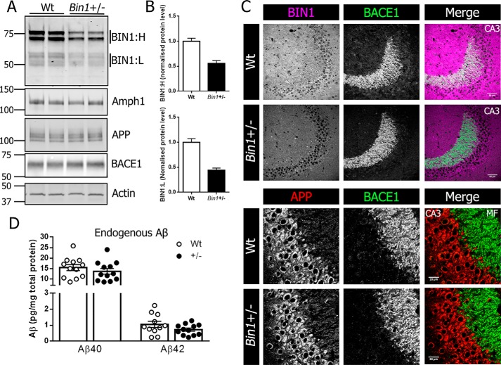
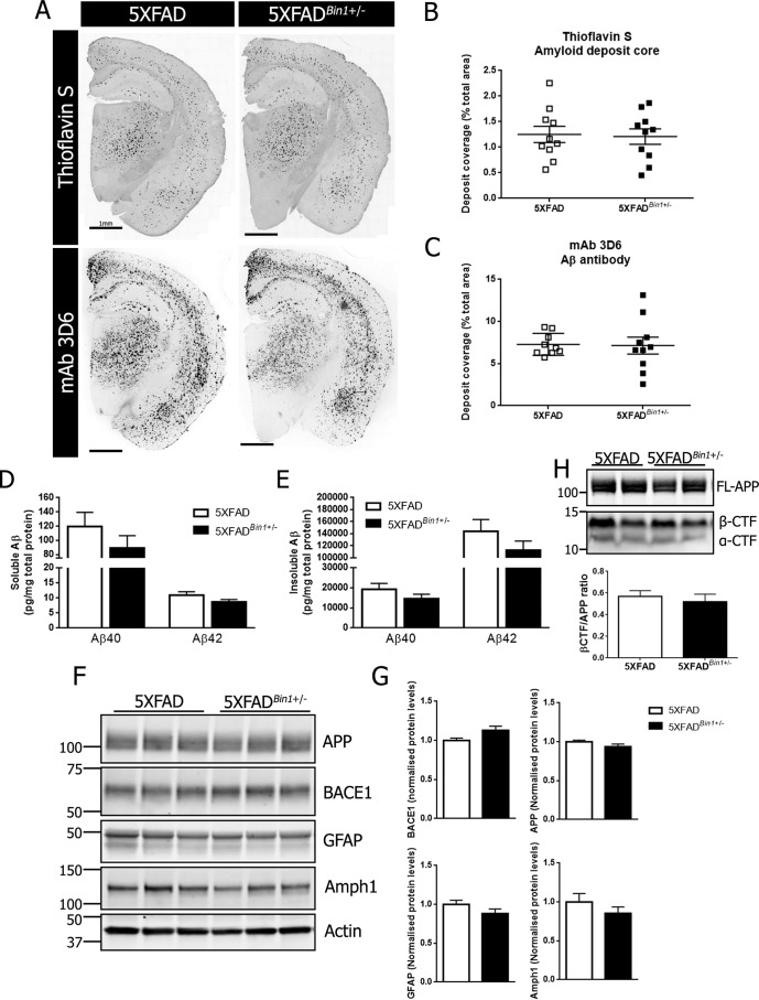
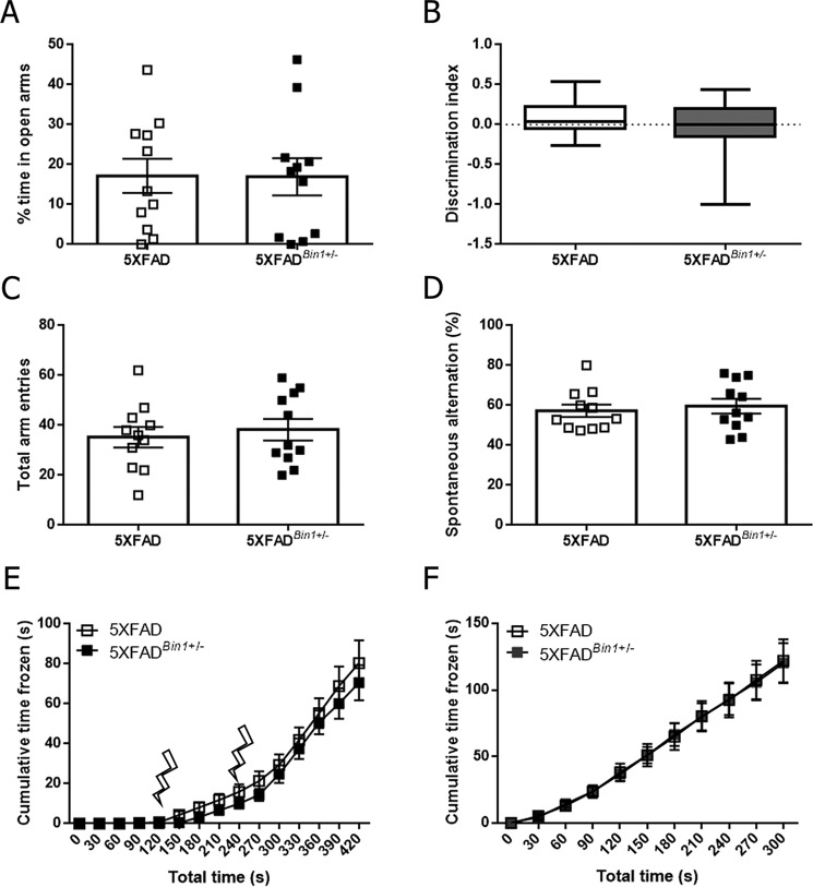
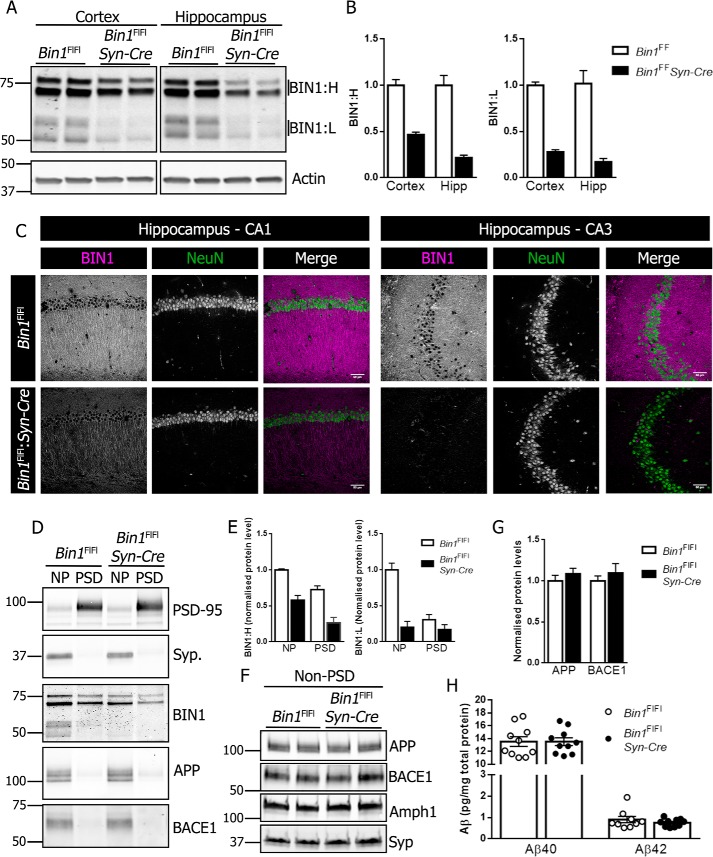
Alzheimer's disease (AD) is pathologically characterized by the deposition of the β-amyloid (Aβ) peptide in senile plaques in the brain, leading to neuronal dysfunction and eventual decline in cognitive function. Genome-wide association studies have identified the bridging integrator 1 (BIN1) gene within the second most significant susceptibility locus for late-onset AD. BIN1 is a member of the amphiphysin family of proteins and has reported roles in the generation of membrane curvature and endocytosis. Endocytic dysfunction is a pathological feature of AD, and endocytosis of the amyloid precursor protein is an important step in its subsequent cleavage by β-secretase (BACE1). In vitro evidence implicates BIN1 in endosomal sorting of BACE1 and Aβ generation in neurons, but a role for BIN1 in this process in vivo is yet to be described. Here, using biochemical and immunohistochemistry analyses we report that a 50% global reduction of BIN1 protein levels resulting from a single Bin1 allele deletion in mice does not change BACE1 levels or localization in vivo, nor does this reduction alter the production of endogenous murine Aβ in nontransgenic mice. Furthermore, we found that reduction of BIN1 levels in the 5XFAD mouse model of amyloidosis does not alter Aβ deposition nor behavioral deficits associated with cerebral amyloid burden. Finally, a conditional BIN1 knockout in excitatory neurons did not alter BACE1, APP, C-terminal fragments derived from BACE1 cleavage of APP, or endogenous Aβ levels. These results indicate that BIN1 function does not regulate Aβ generation in vivo.

Keywords: 5XFAD; APP; Alzheimer disease; BACE1; BIN1; GWAS; amphiphysin; amyloid; amyloid precursor protein (APP); beta-amyloid (AB); beta-secretase 1 (BACE1); endocytosis; neurodegeneration; pathogenesis; secretase.


Conflict of interest statement

The authors declare that they have no conflicts of interest with the contents of this article

Figures

Figure 1.
Figure 1.
Single allele deletion of Bin1 does not alter APP or BACE localization or Aβ levels. A, forebrain homogenates from 4-month-old Bin1+/− mice and WT littermate controls were analyzed by immunoblotting for BIN1, APP, BACE1, and Amph1 levels. B, quantitative analysis of BIN1:H (top) and BIN1:L (bottom) in forebrain lysates from WT and Bin1+/− mice. C, top panel, immunofluorescent staining for BIN1 (magenta) and BACE1 (green) in the hippocampal CA3 region from WT and Bin1+/− mice. Scale bar, 50 μm. Bottom panel, higher magnification of immunofluorescent staining for APP (red) and BACE1 (green) in CA3 neurons and mossy fibers (MF) of the mouse hippocampus. Scale bar, 20 μm. D, forebrain lysates from WT and Bin1+/− mice were analyzed for steady-state levels of endogenous Aβ40 and Aβ42 using a V-PLEX 4G8 immunoassay (Aβ40, n = 12 per genotype; Aβ42, n = 11 WT, and 12 Bin1+/−).
Figure 2.
Figure 2.
Reduction of BIN1 expression does not alter amyloid deposition in female 5XFAD mice. A, representative images of Aβ deposit core staining with thioflavin S and mAb 3D6 immunostaining in 4-month-old female 5XFAD and 5XFADBin1+/− mice at 4 months of age. B, quantification of amyloid burden identified by thioflavin S staining (n = 10 per genotype). C, quantification of amyloid burden identified by mAb 3D6 immunostaining (n = 9 5XFAD and 10 5XFADBin1+/−). Forebrain tissue from 4-month-old female 5XFAD and 5XFADBin1+/− mice was sequentially extracted in TBS and formic acid to generate soluble and insoluble Aβ fractions. D and E, the levels of soluble and insoluble Aβ40 and Aβ42 were measured by V-PLEX 6E10 immunoassay (n = 10 per genotype). F, immunoblot analysis of APP, BACE1, GFAP, and Amph1 levels in 5XFAD and 5XFADBin1+/− mice. G, quantitative analysis of the levels of BACE1, APP, GFAP, and Amph1, normalized to actin (n = 8 per genotype). H, immunoblot analysis of full-length APP (FL-APP) and APP C-terminal fragments (CTF) and quantification of β-CTF normalized to FL-APP (n = 4 per genotype).
Figure 3.
Figure 3.
Reduction in BIN1 expression does not alter behavioral deficits in the absence of changes in amyloid burden in female 5XFAD mice. A, the percentage of time in the open arms of the elevated-plus maze was quantified for 4-month-old 5XFAD and 5XFADBin1+/− mice. B, discrimination index score from novel object recognition for 5XFAD and 5XFADBin1+/− mice. Box plots show the median, maximum, and minimum values. C, the number of arm entries over the 8-min Y-maze trial. D, the number of spontaneous alternations in the Y-maze over the 8-min trial period. E, freezing behavior in female 5XFAD and 5XFADBin1+/− mice during day 1 of fear conditioning. Lightning bolts represent delivery of the shock stimulus. F, freezing behavior in female 5XFAD and 5XFADBin1+/− mice on day 2 of fear conditioning, 24 h after shock application (n = 11 for both genotypes in all analyses).
Figure 4.
Figure 4.
Characterization of neuronal conditional Bin1 knockout. A, brain homogenates from the hippocampus and cortex of 4-month-old Bin1Fl/Fl and Bin1Fl/Fl:Syn-Cre were analyzed for BIN1 expression by immunoblotting. B, quantification of BIN1:H and BIN1:L in the cortex and hippocampus (Hipp) (n = 6 Bin1Fl/Fl and 4 Bin1Fl/Fl:Syn-Cre). C, immunohistochemical analysis of BIN1 and NeuN expression in the CA1 (left panel) and CA3 (right panel) of Bin1Fl/Fl and Bin1Fl/Fl:Syn-Cre mice. D, PSD and non-PSD (NP) membrane fractions were analyzed for PSD-95, synaptophysin, BIN1, APP, and BACE1 by immunoblotting. E, quantitative analysis of BIN1:H and BIN1:L in crude pre-and post-synaptic membranes (n = 4). F, non-PSD membranes were analyzed for APP, BACE1, Amph1, and synaptophysin by immunoblotting. G, quantitative analysis of APP and BACE1 levels in pre-synaptic fractions (n = 4). H, forebrain lysates were analyzed for steady-state levels of Aβ40 and Aβ42 using V-PLEX 4G8 immunoassay (Aβ40, n = 10 per genotype; Aβ42, n = 9 Bin1Fl/Fl and 10 Bin1Fl/Fl:Syn-Cre).

References

    1. Musiek E. S., and Holtzman D. M. (2015) Three dimensions of the amyloid hypothesis: Time, space and “wingmen.” Nat. Neurosci. 18, 800–806 10.1038/nn.4018 - DOI - PMC - PubMed
    1. Harold D., Abraham R., Hollingworth P., Sims R., Gerrish A., Hamshere M. L., Pahwa J. S., Moskvina V., Dowzell K., Williams A., Jones N., Thomas C., Stretton A., Morgan A. R., Lovestone S., et al. (2009) Genome-wide association study identifies variants at CLU and PICALM associated with Alzheimer's disease. Nat. Genet. 41, 1088–1093 10.1038/ng.440 - DOI - PMC - PubMed
    1. Naj A. C., Jun G., Beecham G. W., Wang L.-S., Vardarajan B. N., Buros J., Gallins P. J., Buxbaum J. D., Jarvik G. P., Crane P. K., Larson E. B., Bird T. D., Boeve B. F., Graff-Radford N. R., De Jager P. L., et al. (2011) Common variants at MS4A4/MS4A6E, CD2AP, CD33 and EPHA1 are associated with late-onset Alzheimer's disease. Nat. Genet. 43, 436–441 10.1038/ng.801 - DOI - PMC - PubMed
    1. Seshadri S., Fitzpatrick A. L., Ikram M. A., DeStefano A. L., Gudnason V., Boada M., Bis J. C., Smith A. V., Carassquillo M. M., Lambert J. C., Harold D., Schrijvers E. M., Ramirez-Lorca R., Debette S., Longstreth W. T. Jr., et al. (2010) Genome-wide analysis of genetic loci associated with Alzheimer disease. JAMA 303, 1832–1840 10.1001/jama.2010.574 - DOI - PMC - PubMed
    1. Prokic I., Cowling B. S., and Laporte J. (2014) Amphiphysin 2 (BIN1) in physiology and diseases. J. Mol. Med. 92, 453–463 10.1007/s00109-014-1138-1 - DOI - PubMed

Publication types

MeSH terms

Substances