. 2019 Feb 1;33(3-4):166-179.
doi: 10.1101/gad.319889.118. Epub 2019 Jan 28.

CD44 splice isoform switching determines breast cancer stem cell state

Affiliations

CD44 splice isoform switching determines breast cancer stem cell state

Honghong Zhang et al. Genes Dev. .

Abstract

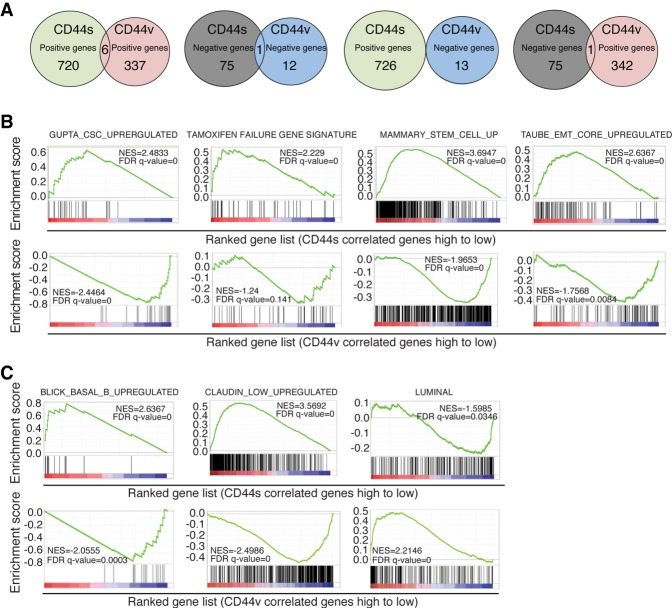
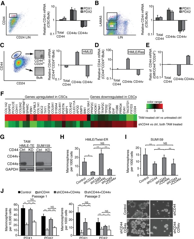
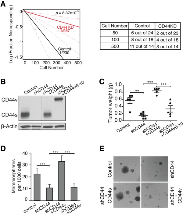
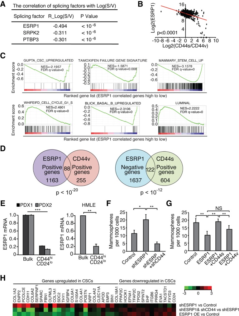
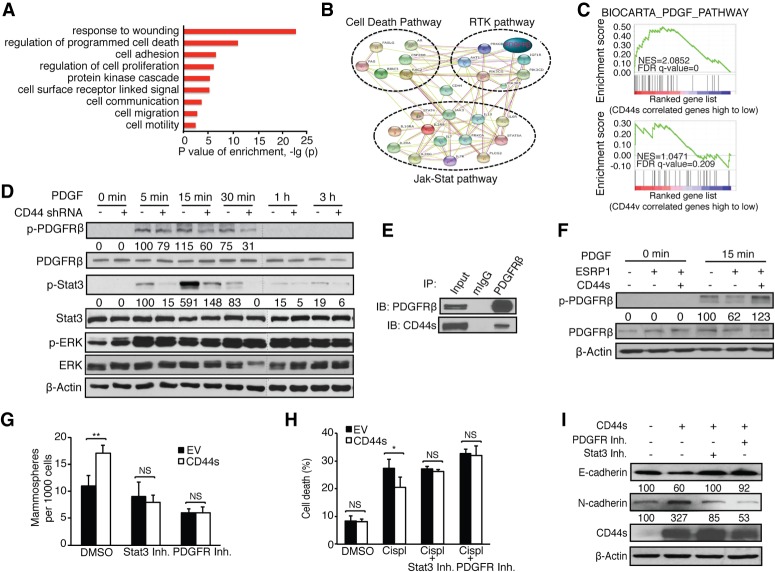
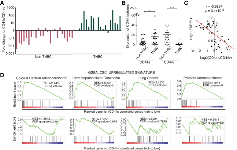
Although changes in alternative splicing have been observed in cancer, their functional contributions still remain largely unclear. Here we report that splice isoforms of the cancer stem cell (CSC) marker CD44 exhibit strikingly opposite functions in breast cancer. Bioinformatic annotation in patient breast cancer in The Cancer Genome Atlas (TCGA) database reveals that the CD44 standard splice isoform (CD44s) positively associates with the CSC gene signatures, whereas the CD44 variant splice isoforms (CD44v) exhibit an inverse association. We show that CD44s is the predominant isoform expressed in breast CSCs. Elimination of the CD44s isoform impairs CSC traits. Conversely, manipulating the splicing regulator ESRP1 to shift alternative splicing from CD44v to CD44s leads to an induction of CSC properties. We further demonstrate that CD44s activates the PDGFRβ/Stat3 cascade to promote CSC traits. These results reveal CD44 isoform specificity in CSC and non-CSC states and suggest that alternative splicing provides functional gene versatility that is essential for distinct cancer cell states and thus cancer phenotypes.

Keywords: CD44s; CSC; PDGFRβ/Stat3; alternative splicing; breast cancer.


Figures

Figure 1.
Figure 1.
Genome-wide TCGA analysis reveals distinct association of CD44 isoforms with breast cancer phenotypes and subtypes. (A) Venn diagram plots showing the overlapping of CD44s- and CD44v-correlated genes analyzed from the breast cancer TCGA database. CD44v levels were calculated as the average of exon expression of v8, v9, and v10. (B) Gene set enrichment analysis (GSEA) showing the enrichment of CSC, tamoxifen failure, mammary stem cell, and EMT gene signatures in the CD44s-correlated gene list and negative enrichment in the CD44v-correlated gene list. (C) GSEA showing the positive enrichment of the Basal_B gene signature and Claudin_low signature in the CD44s-correlated gene list and the Luminal gene signature in the CD44v-correlated gene list.
Figure 2.
Figure 2.
CD44s is the major splice isoform expressed in CSCs and mediates CSC properties. (A) CD44 isoform expression was analyzed in CD44hiCD24lo CSCs and bulk populations that are lineage-negative isolated from two PDX tumors. Distinct primer pairs that amplify all CD44 isoforms (total CD44), CD44s, or v5 and v6 exon-containing CD44v (CD44v5/v6) were used. Ratios of CD44 isoform mRNAs in CSC and bulk populations are shown. (B) CD44 isoform expression was analyzed in LAMA5hi and LAMA5lo populations isolated from two PDX tumors. (C, left panel) The CD44hiCD24lo population was isolated from human mammary epithelial (HMLE) cells by FACS. Inset images show the mammosphere-forming ability of the CD44hiCD24lo population. (Right panel) Ratios of CD44 isoform mRNA levels analyzed in CD44hiCD24lo and bulk HMLE populations are shown. (D) Ratios of CD44 isoform mRNAs in CD44hiCD24lo and bulk HMLE/Ras populations are shown. (E) The FACS-sorted CD44hiCD24lo population of HMLE cells was grown in monolayer culture for 8 d for epithelial differentiation. Ratios of CD44 isoform mRNAs at day 8 relative to day 0 are shown. (F) TAM-treated HMLE/Twist-ER cells expressing control or CD44 shRNA were analyzed by quantitative RT–PCR (qRT–PCR) for expression of the CSC signature. (G) Immunoblot of CD44 isoform expression in TAM-treated HMLE/Twist-ER (HMLE-TE), SUM159, and their corresponding CD44 knockdown cell lines. (HJ) The effect of CD44 isoforms on mammosphere-forming ability was analyzed using breast cell lines (H,I) and PDX-derived tumor cells (J). (J) Representative images of mammospheres are shown in PDX-derived tumor cells with differential CD44 isoform expression. The numbers of mammospheres are presented. Error bars indicate SEM. n = 3. (*) P < 0.05; (**) P < 0.01.
Figure 3.
Figure 3.
CD44s is a functional mediator of tumor-initiating cell properties in a mouse model of breast cancer progression. (A) Recurrent breast tumor cells expressing control or CD44 shRNA (CD44KD) were transplanted into mammary fat pads of FVB mice. Data are presented as a log–log plot. Frequency of CSCs is calculated by extreme limiting dilution analysis. (B) Immunoblot analysis showing reconstituted expression of CD44s and CD44v in CD44 knockdown cells that ectopically expressed CD44s and CD44v cDNA. (C) Analysis of tumor weight in the indicated mice. The CD44v6–10 reconstituted cells formed the least number of tumors; i.e., five tumors from 12 injections. Thus, the five largest tumors from each group were compared. (D) The effect of CD44s in mammosphere-forming ability was assessed in recurrent tumor cells that have had CD44 depleted and its isoforms reconstituted. (E) Representative mammosphere images are shown. Error bars in C and D indicate SEM (n = 5; C) and SD (n = 9; D). (**) P < 0.01; (***) P < 0.001.
Figure 4.
Figure 4.
The splicing factor ESRP1 suppresses CD44s-mediated CSC function and inversely correlates with CSC signatures. (A) The correlation values between splicing factors and the ratio of CD44s to CD44v from analysis of the breast cancer TCGA data set are shown. (B) ESRP1 levels show significant negative correlation with the ratios of CD44s to CD44v. Log2 values are shown. (C) GSEA plots indicate the negative enrichment of CSC, TAM failure, mammary stem cell, and Basal_B gene signatures and positive enrichment of G1–S and Luminal A gene signatures in the ESRP1-correlated gene list. (D) Venn diagram plots showing the overlapping of ESRP1-, CD44v-, and CD44s-associated genes using the breast cancer TCGA data set. (E) qRT–PCR analysis indicates that ESRP1 mRNA levels in the CD44hiCD24lo population was reduced compared with the bulk population in PDX tumors and HMLE cells. (F,G) Mammosphere-forming ability is depicted in TGFβ-treated HMLE cells (control, shESRP1, and shESRP1 + shCD44) or SUM159 cells (control, ESRP1, ESRP1 + CD44s, and ESRP1 + CD44v) after growth in mammosphere culture for 2 wk. (H) HMLE cell lines (control, shESRP1, shESRP1 + shCD44, and ESRP1 overexpression) were analyzed by qRT–PCR for expression of the CSC signature. Error bars in EG indicate SEM. n = 3. (*) P < 0.05; (**) P < 0.01; (***) P < 0.001.
Figure 5.
Figure 5.
CD44s mediates CSC-like properties through the PDGFRβ/Stat3 signaling pathway. (A) Gene ontology analysis of CD44s-correlated genes. (B) STRING analysis showing functional association networks of CD44s-correlated genes. (C) GSEA plots showing the enrichment of the PDGF pathway signature in the CD44s-correlated gene list. (D) The levels of p-PDGFRβ, p-Stat3, and p-ERK were examined in HMLE-Twist control and shCD44 cells using immunoblot analysis. Cells were treated with 10 ng/mL PDGF for the indicated time intervals. The relative intensity of phosphorylated to unphosphorylated signals is depicted below the images. Representative images are shown from three biological replicates. (E) 293FT cells cotransfected with CD44s and Flag-tagged PDGFRβ were subjected to immunoprecipitation (IP). (F) The levels of p-PDGFRβ were examined using immunoblot analysis in HMLE/Twist cells expressing control and ESRP1 cDNA or coexpressing ESRP1 and CD44s cDNA. Cells were treated with 10 ng/mL PDGF for 15 min. The relative intensity of p-PDGFRβ to total PDGFRβ is depicted below the images. Representative images are shown from three biological replicates. (G) Mammosphere-forming ability assay in control and CD44s-expressing Mes10A cells that were treated with 5 µM PDGFR inhibitor IV or 10 µM Stat3 inhibitor XVIII. (H) Mes10A cells expressing control and CD44s cDNA were pretreated with 5 µM PDGFR inhibitor IV or 10 µM Stat3 inhibitor XVIII for 1 h followed by treatment with 100 µM cisplatin (Cispl) for 24 h. Cell death was assessed and plotted as percent dead cells. (I) Mes10A cells expressing CD44s cDNA were treated with 10 µM Stat3 inhibitor XVIII or 5 µM PDGFR inhibitor IV for 48 h. The expression level of E-cadherin and N-cadherin was examined using immunoblot analysis. The parental Mes10A cells were used as control. Representative images are shown from three biological replicates. Error bars in G and H indicate SEM. n = 3. (*) P < 0.05; (**) P < 0.01.
Figure 6.
Figure 6.
CD44s expression is elevated in TNBC and associates with a CSC signature in multiple cancer types. (A) RNA isolated from frozen clinical specimens of TNBC (n = 20) and non-TNBC (n = 24) was subjected to qRT–PCR analysis of CD44s and CD44v. Ratios of CD44s to CD44v are shown. (B) Expression levels of CD44s and CD44v in TNBC and non-TNBC samples are shown. (C) Correlation graphing reveals a significant negative correlation between ESRP1 and the ratio of CD44s to CD44v in breast tumor samples. (D) GSEA plots indicate the enrichment of the CSC gene signature in the CD44s-correlated gene list in colon and rectum adenocarcinoma, liver hepatocellular carcinoma, lung cancer, and prostate adenocarcinoma. Error bars in B indicate SEM. n = 24 non-TNBC; n = 20 TNBC. (*) P < 0.05; (***) P < 0.001.

References

    1. Aceto N, Bardia A, Miyamoto DT, Donaldson MC, Wittner BS, Spencer JA, Yu M, Pely A, Engstrom A, Zhu H, et al. 2014. Circulating tumor cell clusters are oligoclonal precursors of breast cancer metastasis. Cell 158: 1110–1122. 10.1016/j.cell.2014.07.013 - DOI - PMC - PubMed
    1. Al-Hajj M, Wicha MS, Benito-Hernandez A, Morrison SJ, Clarke MF. 2003. Prospective identification of tumorigenic breast cancer cells. Proc Natl Acad Sci 100: 3983–3988. 10.1073/pnas.0530291100 - DOI - PMC - PubMed
    1. Auvinen P, Tammi R, Kosma VM, Sironen R, Soini Y, Mannermaa A, Tumelius R, Uljas E, Tammi M. 2013. Increased hyaluronan content and stromal cell CD44 associate with HER2 positivity and poor prognosis in human breast cancer. Int J Cancer 132: 531–539. 10.1002/ijc.27707 - DOI - PubMed
    1. Bhola NE, Balko JM, Dugger TC, Kuba MG, Sánchez V, Sanders M, Stanford J, Cook RS, Arteaga CL. 2013. TGF-β inhibition enhances chemotherapy action against triple-negative breast cancer. J Clin Invest 123: 1348–1358. 10.1172/JCI65416 - DOI - PMC - PubMed
    1. Brooks MD, Burness ML, Wicha MS. 2015. Therapeutic implications of cellular heterogeneity and plasticity in breast cancer. Cell Stem Cell 17: 260–271. 10.1016/j.stem.2015.08.014 - DOI - PMC - PubMed

Publication types