. 2019 Feb 1;33(3-4):144-149.
doi: 10.1101/gad.321117.118. Epub 2019 Jan 28.

Nuclear pore density controls heterochromatin reorganization during senescence

Affiliations

Nuclear pore density controls heterochromatin reorganization during senescence

Charlene Boumendil et al. Genes Dev. .

Abstract

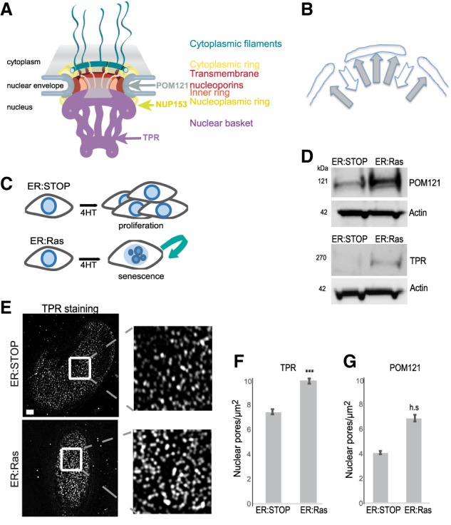
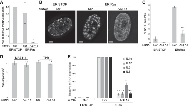
During oncogene-induced senescence (OIS), heterochromatin is lost from the nuclear periphery and forms internal senescence-associated heterochromatin foci (SAHFs). We show that an increased nuclear pore density during OIS is responsible for SAHF formation. In particular, the nucleoporin TPR is necessary for both formation and maintenance of SAHFs. Loss of SAHFs does not affect cell cycle arrest but abrogates the senescence-associated secretory phenotype-a program of inflammatory cytokine gene activation. Our results uncover a previously unknown role of nuclear pores in heterochromatin reorganization in mammalian nuclei and demonstrate the importance of heterochromatin organization for a specific gene activation program.

Keywords: inflammation; nuclear organization; nuclear pore; senescence.


Figures

Figure 1.
Figure 1.
Nuclear pore density increases in OIS. (A) Model of the NPC showing the position of TPR, NUP153, and POM121. (B) Schematic showing the balance of forces attracting heterochromatin to the NL and repelling heterochromatin from nuclear pores. (C) Schematic of OIS induction in ER:Ras cells by 4-hydroxy-tamoxifen (4HT) and continued proliferation in ER:Stop cells. (D) Western blot showing POM121 (left panel) and TPR (right panel) levels in 4HT-treated ER:Stop and ER:Ras cells. (E) TPR immunostaining in ER:Stop and ER:Ras cells treated with 4HT. (Left) The bottom plane of a nucleus imaged by structured illuminated microscopy (SIM). (Right) Enlargement of the insets. Bars, 2 µm. (F) Mean (±SEM) nuclear pore density (pores per square micrometer) in 4HT-treated ER:Stop and ER:Ras cells as counted by TPR staining in three biological replicates. (***) P = 0.0001. (G) As in F, but for Pom121 staining. (h.s.) Highly significant (P = 1.3 × 10−06).
Figure 2.
Figure 2.
Increased nuclear pore density in OIS is necessary for SAHF formation. (A) Schematic showing the depletion experiment for BE. (B) MAB414 (antibody recognizing several nucleoporins) immunostaining in ER:Stop cells treated with 4HT after 2 d of knockdown with scramble (Scr) or POM121 siRNAs. (Left) The bottom plane of the nucleus imaged by SIM. (Right) Enlargement of the insets. Bars, 2 µm. (C) Mean (±SEM) nuclear pore density (pores per square micrometer) in 4HT-treated ER:Stop cells after scramble (Scr) or POM121 siRNA knockdown, as assayed by TPR staining in three biological replicates, (*) P < 0.05. (D) DAPI staining of 4HT-treated ER:Stop and ER:Ras cells in controls (Scr) and upon POM121 depletion (siPOM121). Bars, 10 µm. (Bottom) Enlargement of the insets. (E) Mean (±SEM) percentage of cells containing SAHFs in 4HT-treated ER:Stop and ER:Ras cells after knockdown with scramble (Scr) siRNAs and in 4HT-treated ER:Ras cells with POM121 siRNAs. Data are from three experiments. (*) P < 0.05; (h.s.) highly significant.
Figure 3.
Figure 3.
TPR is necessary for SAHF formation and maintenance. (A) DAPI staining of nonsenescent 4HT-treated ER:Stop and OIS (ER:Ras) cells after control scramble (Scr) siRNA and upon TPR depletion (siTPR). Bars, 10 µm. (B) Mean (±SEM) percentage of cells containing SAHFs in 4HT-treated ER:Stop and ER:Ras cells after knockdown siRNAs as in A. Data are from three experiments. (**) P < 0.01; (h.s.) highly significant. (C) Time course for TPR depletion by siRNA late in the OIS program as performed for DF. (D) DAPI staining of 4HT-treated ER:Stop and OIS cells (ER:Ras) in controls (Scr) and upon TPR depletion (siTPR). Bars, 2 µm. (E) Mean (±SEM) percentage of cells containing SAHFs in 4HT-treated ER:Stop and ER:Ras cells after knockdown with scramble (Scr) siRNAs and in ER:Ras cells with TPR siRNAs. Data are from three experiments. (**) P < 0.01; (h.s.) highly significant. (F) DAPI (blue) and TPR (red) staining of 4HT-treated ER:Stop and ER:Ras upon TPR depletion (siTPR) imaged by SIM. (Right) Enlargement of the insets. Bars, 2 µm. (G, top) DAPI (blue) and TPR (red) staining of 4HT-treated ER:Ras cells upon TPR depletion. (Bottom) Costaining with the nucleoporin antibody MAB414 (green).
Figure 4.
Figure 4.
TPR is necessary for the SASP. (A) Mean (±SEM) mRNA level measured by quantitative RT–PCR (qRT–PCR) for SASP genes (IL1A, IL1B, IL6, and IL8) in 4HT-treated ER:Stop and ER:Ras cells after knockdown with scramble (Scr) siRNAs and in 4HT-treated ER:Ras cells with TPR siRNAs. Expression is relative to ER:Ras cells transfected with Scr siRNAs. Data are from three experiments. (h.s.) Highly significant. (B) Mean (±SEM) percentage of cells positive by immunostaining for SASP cytokines (IL1α, IL1β, IL6, and IL8) in 4HT-treated ER:Stop and ER:Ras cells after siRNA knockdown as in A. Data are from three experiments. (**) P < 0.01; (***) P < 0.001; (h.s.) highly significant. (C) Immunostaining (green) for IL1α and IL1β in DAPI-stained (blue) nuclei of 4HT-treated ER:Stop and ER:Ras cells subjected to RNAi as in A. Bars, 100 µm.
Figure 5.
Figure 5.
Chromatin reorganization seems necessary for the SASP. (A) Mean (±SEM) ASF1a mRNA level established by qRT–PCR in 4HT-treated ER:Stop and ER:Ras cells after knockdown with scramble (Scr) or ASF1a siRNAs. Expression is shown relative to ER:Stop cells transfected with Scr siRNAs. Data are from three experiments. (*) P < 0.05. (B) DAPI staining of 4HT-treated ER:Stop and ER:Ras cells in controls (Scr) and upon ASF1a depletion (siASF1a). Bars, 2 µm. (C) Mean (±SEM) percentage of cells containing SAHFs in 4HT-treated ER:Stop and ER:Ras cells after knockdown with scramble (Scr) siRNAs and in ER:Ras cells with ASF1a siRNAs. Data are from three experiments. (***) P < 0.001. (D) Mean (±SEM) nuclear pore density (pores per square micrometer) in 4HT-treated ER:Stop cells after knockdown with scramble (Scr) or ASF1a (siASF1a) siRNAs as counted by MAB414 or TPR staining in three biological replicates. (n.s.) Nonsignificant. (E) Mean (±SEM) mRNA levels measured by qRT–PCR for IL1A, IL1B, IL6, and IL8 in 4HT-treated ER:Stop and ER:Ras cells after knockdown with scramble (Scr) siRNAs and in 4HT-treated ER:Ras cells with ASF1a siRNAs. Expression is shown relative to ER:Ras cells transfected with Scr siRNAs. Data are from three experiments. (***) P < 0.001; (h.s.) highly significant.

Comment in

  • The unusual SASPects.
    Strzyz P. Strzyz P. Nat Rev Mol Cell Biol. 2019 Apr;20(4):195. doi: 10.1038/s41580-019-0111-9. Nat Rev Mol Cell Biol. 2019. PMID: 30770903 No abstract available.

References

    1. Acosta JC, O'Loghlen A, Banito A, Guijarro MV, Augert A, Raguz S, Fumagalli M, Da Costa M, Brown C, Popov N, et al. 2008. Chemokine signaling via the CXCR2 receptor reinforces senescence. Cell 133: 1006–1018. 10.1016/j.cell.2008.03.038 - DOI - PubMed
    1. Acosta JC, Banito A, Wuestefeld T, Georgilis A, Janich P, Morton JP, Athineos D, Kang T-W, Lasitschka F, Andrulis M, et al. 2013. A complex secretory program orchestrated by the inflammasome controls paracrine senescence. Nat Cell Biol 15: 978–990. 10.1038/ncb2784 - DOI - PMC - PubMed
    1. Bodoor K, Shaikh S, Salina D, Raharjo WH, Bastos R, Lohka M, Burke B. 1999. Sequential recruitment of NPC proteins to the nuclear periphery at the end of mitosis. J Cell Sci 112: 2253–2264. - PubMed
    1. Capelson M, Hetzer MW. 2009. The role of nuclear pores in gene regulation, development and disease. EMBO Rep 10: 697–705. 10.1038/embor.2009.147 - DOI - PMC - PubMed
    1. Chandra T, Kirschner K, Thuret J-Y, Pope BD, Ryba T, Newman S, Ahmed K, Samarajiwa SA, Salama R, Carroll T, et al. 2012. Independence of repressive histone marks and chromatin compaction during senescent heterochromatic layer formation. Mol Cell 47: 203–214. 10.1016/j.molcel.2012.06.010 - DOI - PMC - PubMed

Publication types

MeSH terms