. 2019 Feb 1;33(3-4):209-220.
doi: 10.1101/gad.317412.118. Epub 2019 Jan 28.

A ZNRF3-dependent Wnt/β-catenin signaling gradient is required for adrenal homeostasis

Affiliations

A ZNRF3-dependent Wnt/β-catenin signaling gradient is required for adrenal homeostasis

Kaitlin J Basham et al. Genes Dev. .

Abstract

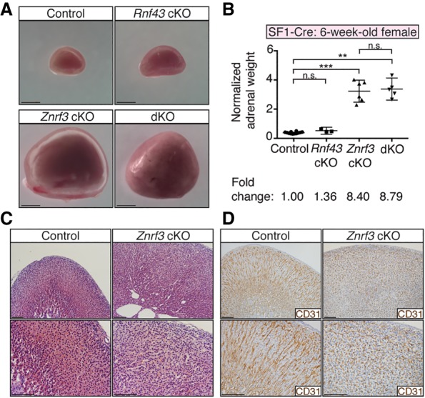
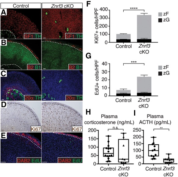
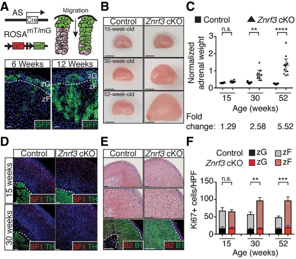
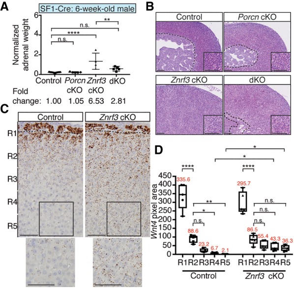
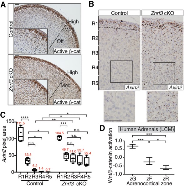
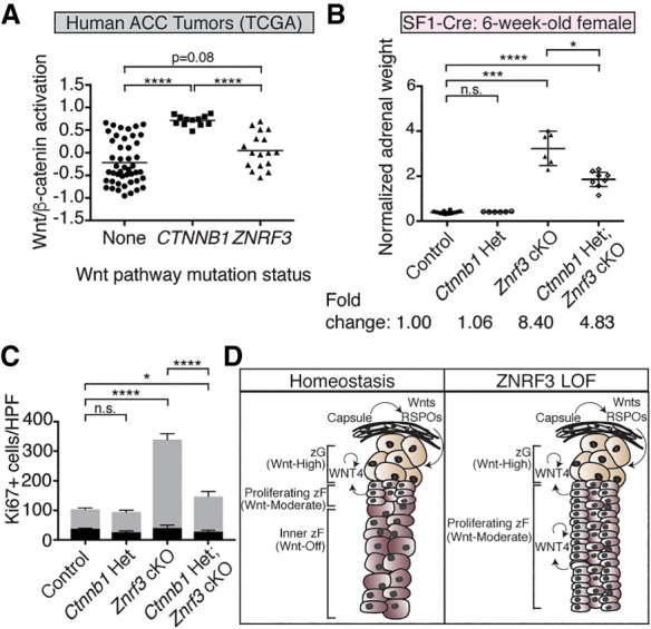
Spatiotemporal control of Wnt signaling is essential for the development and homeostasis of many tissues. The transmembrane E3 ubiquitin ligases ZNRF3 (zinc and ring finger 3) and RNF43 (ring finger protein 43) antagonize Wnt signaling by promoting degradation of frizzled receptors. ZNRF3 and RNF43 are frequently inactivated in human cancer, but the molecular and therapeutic implications remain unclear. Here, we demonstrate that adrenocortical-specific loss of ZNRF3, but not RNF43, results in adrenal hyperplasia that depends on Porcupine-mediated Wnt ligand secretion. Furthermore, we discovered a Wnt/β-catenin signaling gradient in the adrenal cortex that is disrupted upon loss of ZNRF3. Unlike β-catenin gain-of-function models, which induce high Wnt/β-catenin activation and expansion of the peripheral cortex, ZNRF3 loss triggers activation of moderate-level Wnt/β-catenin signaling that drives proliferative expansion of only the histologically and functionally distinct inner cortex. Genetically reducing β-catenin dosage significantly reverses the ZNRF3-deficient phenotype. Thus, homeostatic maintenance of the adrenal cortex is dependent on varying levels of Wnt/β-catenin activation, which is regulated by ZNRF3.

Keywords: Wnt signaling; adrenal zonation; mouse models; organ maintenance; proliferation.


Figures

Figure 1.
Figure 1.
ZNRF3 is expressed throughout the adrenal cortex, beneath the RSPO-producing capsule. (A,B) Rnf43 is expressed predominately in the zG (A), while Znrf3 is expressed throughout the cortex (B). The dashed line marks the histological zG/zF boundary. Bars, 50 µm. Representative images of single-molecule in situ hybridizations (ISHs) from 6-wk-old female mice are shown. (C,D) Quantification of ISHs based on pixel area. Statistical analysis was performed using two-tailed Student's t-test. (*) P < 0.05.
Figure 2.
Figure 2.
Loss of ZNRF3 induces rapid adrenal growth. (A) Whole adrenals from Znrf3 cKO and Rnf43;Znrf3 double-knockout (dKO) mice are significantly larger in size at 6 wk compared with control or Rnf43 cKO mice. Bars, 1 mm. (B) Normalized adrenal weights shown as mean and 95% confidence interval (CI). Statistical analysis was performed using Welch's one-way ANOVA followed by Games-Howell post hoc test. (**) P < 0.01; (***) P < 0.001. (C,D) Loss of ZNRF3 disrupts normal adrenocortical architecture, as shown by H&E (C) and IHC (D) for the vascular marker CD31. Bars, 100 µm. All data shown are from female mice.
Figure 3.
Figure 3.
Loss of ZNRF3 results in proliferative expansion of the zF. (AC) ZNRF3 loss expands the zF and disrupts organization of the inner adrenal medulla. Dashed lines mark histological cortical/medullary and zG/zF boundaries. SF1 (cortex), TH (medulla), B2 (zG), B1 (zF), and 20α-HSD (X zone) were used as markers. (D,E) ZNRF3 loss significantly increases proliferation in the zF based on IHC for Ki67 (D) and EdU (E) incorporation. DAB2 marks the zG. (F,G) Quantification of Ki67 and EdU based on the number of positive cells per high-power field (HPF) within the histological zG or zF. Results are shown as mean ± SEM with three biological replicates per genotype. Statistical analysis was performed using two-way ANOVA followed by Tukey's post hoc test. (***) P < 0.001; (****) P < 0.0001. Reported statistical results represent comparison between zF compartments (gray). No significant difference was observed between zG compartments (black). (H,I) Znrf3 cKO mice maintain normal plasma corticosterone concentrations (H) concomitant with decreased adrenocorticotropic hormone (ACTH) (I). Statistical analysis was performed using two-tailed Welch's t-test. (**) P < 0.01. All data shown are from female mice. Bars, 50 µm.
Figure 4.
Figure 4.
AS-Cre recapitulates SF1-Cre-driven ZNRF3 loss, supporting an adrenal-specific defect in the zF. (A) AS-Cre expression begins in the zG, which gives rise to the zF. Bars, 20 µm. (B) Adrenal glands from AS-Cre-driven Znrf3 cKO mice are significantly larger in size by 30 wk compared with controls. Bars, 1 mm. (C) Normalized adrenal weights are shown as mean and 95% CI. Statistical analysis was performed using two-tailed Welch's t-test. (**) P < 0.01; (****) P < 0.0001. (D,E) AS-Cre-driven loss of ZNRF3 recapitulates the phenotype observed with SF1-Cre, including progressive disruption of the innermost TH-expressing medulla by SF1-positive (D) and CYP11B1-positive (E) zF cells. Bars, 100 µm. (F) AS-Cre-driven loss of ZNRF3 significantly increases proliferation. Quantification of Ki67 based on the number of positive cells per high-power field (HPF) within the histological zG or zF. Results are shown as mean ± SEM with at least three biological replicates per genotype. Statistical analysis was performed using two-way ANOVA followed by Tukey's post hoc test. (**) P < 0.01; (***) P < 0.001. Reported statistical results represent comparison between zF compartments (dashed line). No significant difference was observed between zG compartments (solid line). Dashed lines mark histological cortical/medullary and zG/zF boundaries. All data shown are from female mice.
Figure 5.
Figure 5.
Manifestation of the ZNRF3-deficient phenotype requires Wnt ligand secretion. (A,B) Loss of PORCN significantly rescues the increase in adrenal weight (A) and disrupted adrenocortical architecture (B) observed with ZNRF3 loss. (dKO) Double knockout. Normalized adrenal weight is shown as mean and 95% CI. Statistical analysis was performed using one-way ANOVA followed by Tukey's post hoc test. (**) P < 0.01; (****) P < 0.0001. Representative H&Es from male cKOs are shown. The dashed line marks the histological cortical/medullary boundary. Insets show zF. Bars, 100 µm. (C) Wnt4, which is expressed along a gradient in the normal adrenal cortex, is significantly increased in the inner cortex of Znrf3 cKOs. Single-molecule ISHs were performed in 6-wk-old females. The dashed line marks the histological zG/zF boundary. Bars, 50 µm. (D) Quantification of Wnt4 ISH data based on pixel area within five equal-sized regions (R1–R5) extending from the outer capsule. Results are shown as mean ± SEM with five biological replicates per genotype. The mean pixel area for each region is noted in red. Statistical analysis was performed using one-way ANOVA followed by Tukey's post hoc test. (*) P < 0.05; (**) P < 0.01; (****) P < 0.0001.
Figure 6.
Figure 6.
Loss of ZNRF3 increases moderate-level Wnt/β-catenin signaling to promote adrenal hyperplasia. (AC) ZNRF3 loss increases activated β-catenin (A) and Axin2 (B,C) expression in the zF. Representative images from 6-wk-old females are shown. The dashed line marks the histological zG/zF boundary. Bars, 50 µm. Axin2 quantification based on pixel area within five equal-sized regions (R1–R5) extending from the outer capsule. Results are shown as mean ± SEM with four biological replicates per genotype. The mean pixel area for each region is noted in red. Statistical analysis was performed using one-way ANOVA followed by Tukey's post hoc test. (*) P < 0.05; (**) P < 0.01; (***) P < 0.001; (****) P < 0.0001. (D) Wnt/β-catenin activation follows a gradient in the normal human adrenal cortex. Each zone was isolated by laser capture microdissection (LCM). Statistical analysis was performed using one-way ANOVA followed by Tukey's post hoc test. (*) P < 0.05; (***) P < 0.001.
Figure 7.
Figure 7.
Reduced β-catenin dosage significantly reverses the ZNRF3-deficient phenotype. (A) Wnt/β-catenin activation in ZNRF3-altered human ACCs is significantly lower compared with CTNNB1-activated tumors. Statistical analysis was performed using Welch's one-way ANOVA followed by Games-Howell post hoc test. (****) P < 0.0001. (B,C) Loss of a single copy of Ctnnb1 in the context of ZNRF3 loss significantly rescues adrenal weight (B) and proliferation (C). Normalized adrenal weights are shown as mean and 95% CI. Statistical analysis was performed using Welch's one-way ANOVA followed by Games-Howell post hoc test. (*) P < 0.05; (***) P < 0.001; (****) P < 0.0001. Quantification of Ki67 based on the number of positive cells per high-power field (HPF) within the histological zG or zF. Results are shown as mean ± SEM with three biological replicates per genotype. Statistical analysis was performed using two-way ANOVA followed by Tukey's post hoc test. (*) P < 0.05; (****) P < 0.0001. Reported statistical results represent comparison between zF compartments (gray). No significant difference was observed between zG compartments (black). (D) Model for how varying levels of Wnt/β-catenin signaling regulate adrenal homeostasis. High Wnt/β-catenin signaling in the zG, which is driven in part by capsular-derived Wnts and RSPOs, helps promote zG differentiation. As zG cells migrate centripetally, moderate-level Wnt/β-catenin signaling supports proliferation and conversion into zF. Wnt/β-catenin signaling is ultimately shut off in the innermost cortex to facilitate proper homeostatic turnover. With loss of ZNRF3, moderate-level Wnt/β-catenin signaling is sustained throughout the inner cortex, resulting in increased proliferation and hyperplasia.

References

    1. Åkerström T, Maharjan R, Sven Willenberg H, Cupisti K, Ip J, Moser A, Stalberg P, Robinson B, Alexander Iwen K, Dralle H, et al. 2016. Activating mutations in CTNNB1 in aldosterone producing adenomas. Sci Rep 6: 19546 10.1038/srep19546 - DOI - PMC - PubMed
    1. Assie G, Letouze E, Fassnacht M, Jouinot A, Luscap W, Barreau O, Omeiri H, Rodriguez S, Perlemoine K, Rene-Corail F, et al. 2014. Integrated genomic characterization of adrenocortical carcinoma. Nat Genet 46: 607–612. 10.1038/ng.2953 - DOI - PubMed
    1. Barrott JJ, Cash GM, Smith AP, Barrow JR, Murtaugh LC. 2011. Deletion of mouse Porcn blocks Wnt ligand secretion and reveals an ectodermal etiology of human focal dermal hypoplasia/Goltz syndrome. Proc Natl Acad Sci 108: 12752–12757. 10.1073/pnas.1006437108 - DOI - PMC - PubMed
    1. Berthon A, Sahut-Barnola I, Lambert-Langlais S, de Joussineau C, Damon-Soubeyrand C, Louiset E, Taketo MM, Tissier F, Bertherat J, Lefrancois-Martinez AM, et al. 2010. Constitutive β-catenin activation induces adrenal hyperplasia and promotes adrenal cancer development. Hum Mol Genet 19: 1561–1576. 10.1093/hmg/ddq029 - DOI - PubMed
    1. Berthon A, Drelon C, Ragazzon B, Boulkroun S, Tissier F, Amar L, Samson-Couterie B, Zennaro MC, Plouin PF, Skah S, et al. 2014. WNT/β-catenin signalling is activated in aldosterone-producing adenomas and controls aldosterone production. Hum Mol Genet 23: 889–905. 10.1093/hmg/ddt484 - DOI - PubMed

Publication types

LinkOut - more resources