. 2019 Mar 27;39(13):2369-2382.
doi: 10.1523/JNEUROSCI.1661-18.2019. Epub 2019 Jan 28.

Lactate Mediates the Effects of Exercise on Learning and Memory through SIRT1-Dependent Activation of Hippocampal Brain-Derived Neurotrophic Factor (BDNF)

Affiliations

Lactate Mediates the Effects of Exercise on Learning and Memory through SIRT1-Dependent Activation of Hippocampal Brain-Derived Neurotrophic Factor (BDNF)

Lauretta El Hayek et al. J Neurosci. .

Abstract

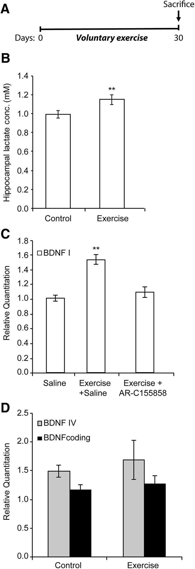
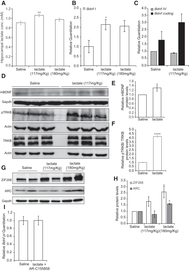
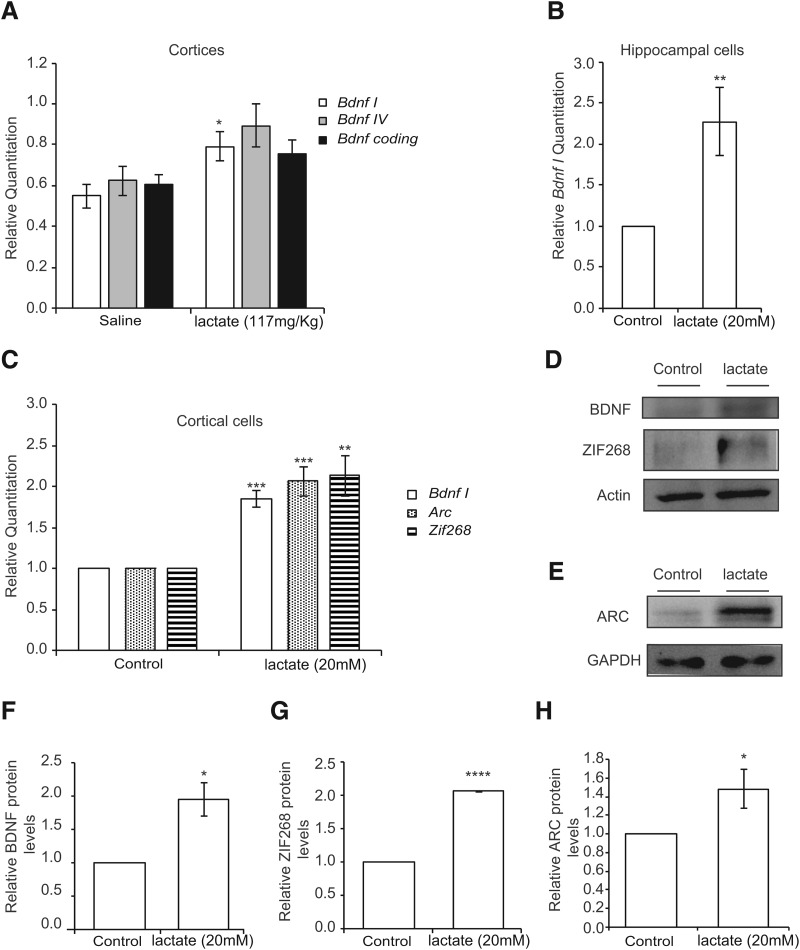
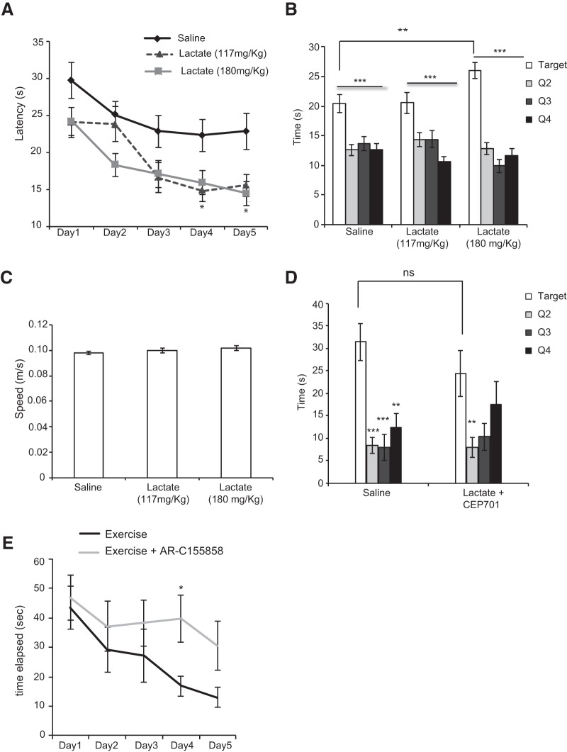
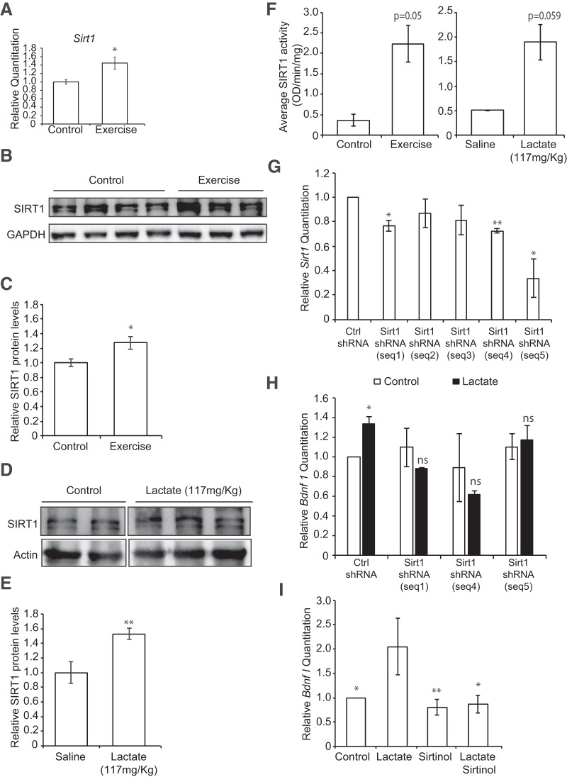
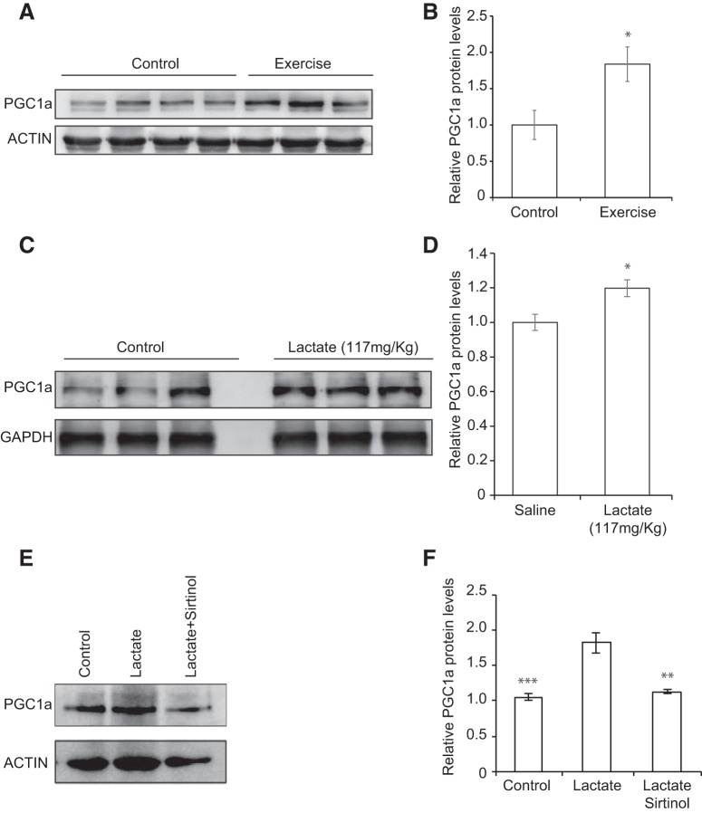
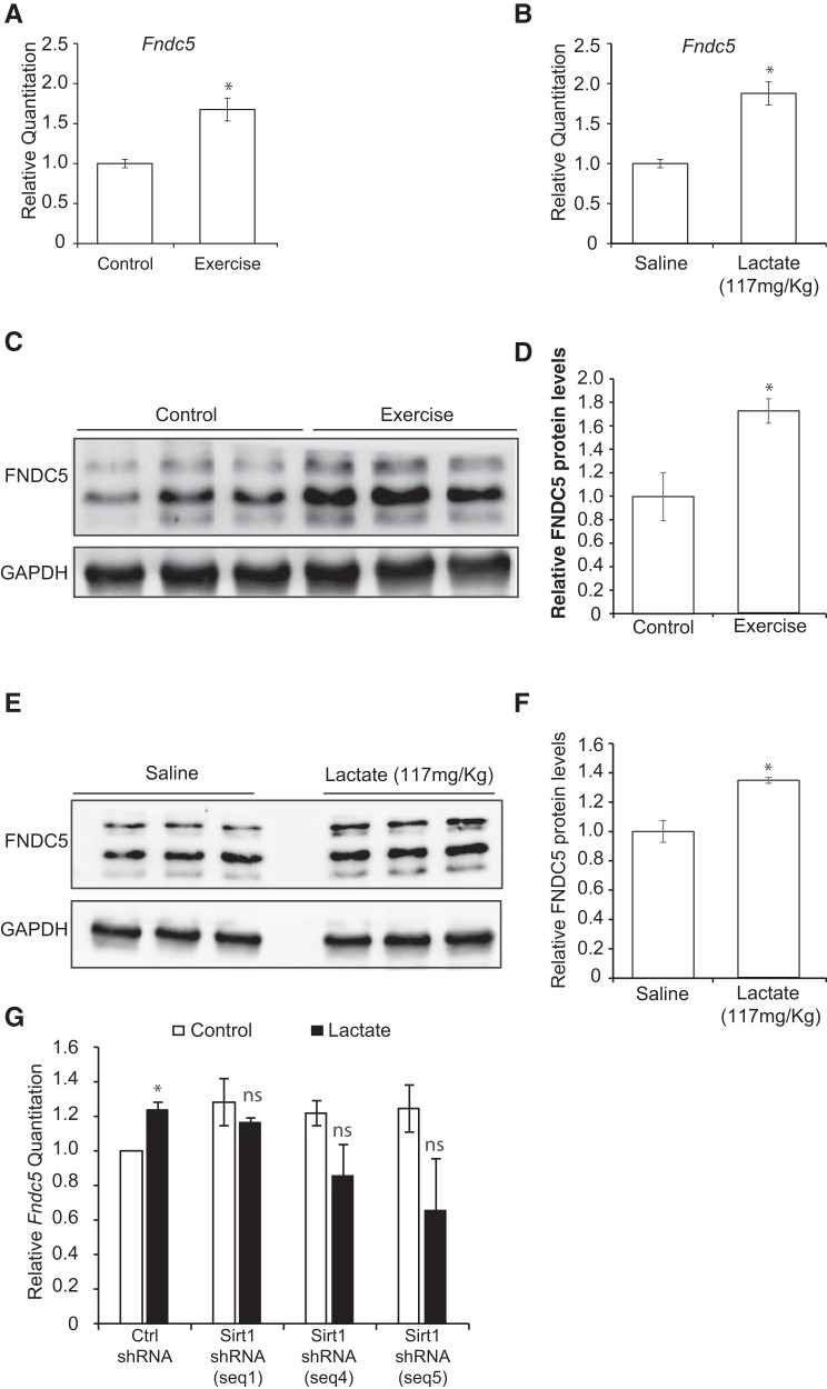
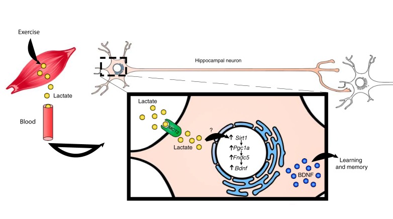
Exercise promotes learning and memory formation. These effects depend on increases in hippocampal BDNF, a growth factor associated with cognitive improvement and the alleviation of depression symptoms. Identifying molecules that are produced during exercise and that mediate hippocampal Bdnf expression will allow us to harness the therapeutic potential of exercise. Here, we report that an endogenous molecule produced during exercise in male mice induces the Mus musculus Bdnf gene and promotes learning and memory formation. The metabolite lactate, which is released during exercise by the muscles, crosses the blood-brain barrier and induces Bdnf expression and TRKB signaling in the hippocampus. Indeed, we find that lactate-dependent increases in BDNF are associated with improved spatial learning and memory retention. The action of lactate is dependent on the activation of the Sirtuin1 deacetylase. SIRT1 increases the levels of the transcriptional coactivator PGC1a and the secreted molecule FNDC5, known to mediate Bdnf expression. These results reveal an endogenous mechanism to explain how physical exercise leads to the induction of BDNF, and identify lactate as a potential endogenous molecule that may have therapeutic value for CNS diseases in which BDNF signaling is disrupted.SIGNIFICANCE STATEMENT It is established that exercise promotes learning and memory formation and alleviates the symptoms of depression. These effects are mediated through inducing Bdnf expression and signaling in the hippocampus. Understanding how exercise induces Bdnf and identifying the molecules that mediate this induction will allow us to design therapeutic strategies that can mimic the effects of exercise on the brain, especially for patients with CNS disorders characterized by a decrease in Bdnf expression and who cannot exercise because of their conditions. We identify lactate as an endogenous metabolite that is produced during exercise, crosses the blood-brain barrier and promotes hippocampal dependent learning and memory in a BDNF-dependent manner. Our work identifies lactate as a component of the "exercise pill."

Keywords: BDNF; PGC1a; Sirt1; exercise; learning; memory formation.


Figures

Figure 1.
Figure 1.
Lactate mediates in part the voluntary exercise-mediated induction of hippocampal Bdnf expression and promotion of learning. A, The exercise paradigm involves 4 weeks of voluntary exercise followed by animal death and hippocampal isolation. B, Voluntary exercise for 4 weeks significantly increases hippocampal lactate levels. The number of hippocampi used for each group (control and exercise) is 10 and 9, respectively. **p < 0.01 (unpaired t test; p = 0.0366 and df = 17). C, Voluntary exercise significantly induces Bdnf promoter I expression in the hippocampus as measured by real-time RT-PCR, whereas inhibiting the lactate MCT transporters by AR-C155858 (50 nm/mouse) abolishes this induction. The number of hippocampi used for each group (control, exercise, and exercise + AR-C155858) is 5, 2, and 4, respectively.**p < 0.01 (one-way ANOVA followed by Dunnett's multiple-comparison test; p = 0.001 for exercise vs control and p = 0.4778 for exercise + AR-C155858 vs control). D, Voluntary exercise does not induce Bdnf pIV or coding expression in the hippocampus as measured by real-time RT-PCR.
Figure 2.
Figure 2.
Peripheral delivery of lactate induces hippocampal Bdnf expression and signaling. A, Intraperitoneal injections of lactate to reach equivalent levels to those reported in the blood after exercise lead to increases in hippocampal lactate. This increase is equivalent to the increase in hippocampal lactate observed after exercise. The number of hippocampi used for each group (control, lactate 117 mg/kg, or lactate 180 mg/kg) is 10, 9, and 7, respectively. **p < 0.01 (one-way ANOVA followed by the Dunnett's post-test; p = 0.003 for lactate 117 mg/kg vs control). B, Intraperitoneal injection of lactate (117 or 180 mg/kg) similar to levels reported during exercise significantly induces hippocampal Bdnf pI expression levels as measured by real-time RT-PCR. The expression was analyzed from the hippocampi of 5 control mice, 5 mice receiving 117 mg/kg lactate, and 5 mice receiving 180 mg/kg lactate. *p < 0.05 (one-way ANOVA followed by the Dunnett's multiple-comparison test; p = 0.0316 for lactate 117 mg/kg vs control and p = 0.0564 for lactate 180 mg/kg vs control). C, Intraperitoneal injection of lactate (117 mg/kg) does not induce hippocampal Bdnf pIV and coding expression levels as measured by real-time RT-PCR. The expression was analyzed from the hippocampi of 4 control mice and 4 mice receiving 117 mg/kg lactate. D, Representative Western blot image depicting the increase in BDNF protein levels and in phosphorylation in the BDNF receptor TRKBB (pTRKB) in the hippocampus of control animals compared with mice receiving 117 mg/kg of lactate. E, Quantification of the BDNF Western blot. *p < 0.05 (unpaired t test; p = 0.043 and df = 4). F, Quantification of the pTRKB Western blot. ****p < 0.0001 (unpaired t test; p = 0.000052 and df = 4). G, Representative Western blot image depicting the increase after 10 min in ZIF268 and ARC protein levels in the hippocampus of control animals compared with mice receiving 117 and 180 mg/kg of lactate. H, Quantification of the ZIF268 and ARC Western blots. *p < 0.05 (one-way ANOVA followed by the Dunnett's multiple-comparison test; ZIF268, p = 0.0196, df = 9 for lactate 180 mg/kg vs control; ARC, p = 0.0303, df = 7 for lactate 180 mg/kg vs control). I, Intraperitoneal injection of lactate (180 mg/kg) along with the lactate MCT transporters AR-C 155858 (50 nm/mouse) did not induce hippocampal Bdnf pI expression levels as measured by real-time RT-PCR. The expression was analyzed from the hippocampi of 5 control mice, 5 mice receiving 180 mg/kg lactate, and AR-C 155858 (50 nm/mouse). Significance was measured by unpaired t test (p = 0.9910 and df = 8).
Figure 3.
Figure 3.
Lactate induces exercise-dependent Bdnf pI and activity-dependent gene expression in mouse primary neurons. A, Intraperitoneal injection of lactate (117 mg/kg) significantly induces cortical Bdnf pI but not pIV or coding expression levels as measured by real-time RT-PCR. The expression was analyzed from the hippocampi of 4 control mice, 4 mice receiving 117 mg/kg lactate. *p < 0.05 (unpaired t test; p = 0.036, df = 6 for lactate 117 mg/kg vs control). B, Lactate significantly induces Bdnf pI expression in primary hippocampal neurons as measured by real-time RT-PCR. **p < 0.01 (unpaired t test; p = 0.0073 and df = 9 for lactate 20 mm vs control). n = 5. Each replicate consisted of primary neurons obtained from different cultures and treated with fresh dilutions of the compounds for 1 h. C, Lactate significantly induces Bdnf pI and activity-dependent gene (Arc and Zif268) expression in primary cortical neurons as measured by real-time RT-PCR. **p < 0.01 (unpaired t test; Bdnf pI, p = 0.0014 and df = 4 for lactate 20 mm vs control; Arc, p = 0.0014 and df = 5 for lactate 20 mm vs control; Zif268, p = 0.0039 and df = 6 for lactate 20 mm vs control). n = 4. Each replicate consisted of primary neurons obtained from different cultures and treated with fresh dilutions of the compounds for 1 h. D, E, Representative Western blot images depicting the lactate-mediated increase in BDNF, ZIF268, and ARC protein levels in mixed (cortical and hippocampal) primary neurons. F, Quantification of the BDNF Western blots. *p < 0.05 (unpaired t test; p = 0.0199, df = 4). G, Quantification of the ZIF268 Western blots. *p < 0.05 (unpaired t test; p < 0.0001, df = 2). H, Quantification of the ARC Western blots. *p < 0.05 (unpaired t test; p = 0.0483, df = 4). ***p < 0.001; ****p < 0.0001.
Figure 4.
Figure 4.
Peripheral delivery of lactate promotes learning and memory. A, Animals were trained for 5 d in a spatial version of the MWM. Animals receiving intraperitoneal injections of lactate (118 and 180 mg/kg) showed significantly reduced escape latency or the time (seconds) required to escape. Results are expressed as mean ± SEM. *p < 0.05 (two-way ANOVA followed by the Bonferroni post-test). B, Animals receiving intraperitoneal injections of lactate (180 mg/kg) spent significantly more time in the target quadrant during a 60 s probe test performed 1 d after the last training session. Results are expressed as mean ± SEM. Statistical significance between the target and the other three quadrants: ***p < 0.0001 (one-way ANOVA followed by Dunnett's post-test). Statistical significance between the target quadrants in saline and lactate-receiving mice: unpaired t test (p = 0.0056 and df = 65). C, The swimming speeds (meter/seconds) of the animals receiving intraperitoneal injections lactate were similar to those receiving saline, indicating that the effects observed in the training and probe test phase were not due to differences in motor behavior between the two groups of animals. D, Animals receiving intraperitoneal injections of lactate (180 mg/kg) in combination with the TRK inhibitor, CEP 701 (3 mg/kg), did not spend significantly more time in the target quadrant compared with animals receiving intraperitoneal injections of saline during a 60 s probe test performed 1 d after the last training session. Results are expressed as mean ± SEM. The number of animals used in each group is 5. Statistical significance was measured by the unpaired t test (p = 0.3117, df = 8). As expected, animals receiving saline spend significantly more time in the target quadrant. Statistical significance was measured by one-way ANOVA followed by the Dunnett's post-test; p = 0.0002 for target versus Quadrant 2 (Q2) and 3 (Q3) and p = 0.0014 for target versus Quadrant 4 (Q4). In contrast, animals receiving lactate and TRK inhibitor CEP701 did not spend significantly more time in the target quadrant compared with Q3 and Q4. One-way ANOVA followed by the Dunnett's post-test; p = 0.0295 for target versus Q2, p = 0.0652 for target versus Q3, and p = 0.4912 for target versus Q4. E, Inhibition of the lactate MCT transporters abolishes exercise-mediated spatial learning in the MWM paradigm. Exercise animals receiving intraperitoneal injections of the lactate MCT transporter inhibitor, AR-C155858 (50 nm/mouse) showed significantly increased escape latency or the time (seconds) required to escape compared with exercise animals receiving intraperitoneal injections of saline. Results are expressed as mean ± SEM. *p < 0.05 (two-way ANOVA followed by the Bonferroni post-test). **p < 0.01.
Figure 5.
Figure 5.
Lactate induces Bdnf expression in an SIRT1-dependent manner. A, Voluntary exercise for 4 weeks significantly induced Sirt1 expression in the hippocampus as measured by real-time RT-PCR. The number of animals used for each group (control and exercise) is 3 and 5, respectively. *p < 0.05 (unpaired t test; p = 0.026 and df = 6). B, Representative Western blot image depicting the increase in SIRT1 protein levels in the hippocampus of control animals compared with exercising mice. C, Quantification of the SIRT1 Western blot. For control and exercise, n = 4. *p < 0.05 (unpaired t test; p = 0.0456 and df = 6). D, Representative Western blot image depicting the increase in SIRT1 protein levels in the hippocampus of mice receiving 117 mg/kg of lactate compared with mice receiving saline. E, Quantification of the SIRT1 Western blot. n = 5 saline injections; n = 6 lactate injections. **p < 0.005 (unpaired t test; p = 0.0077 and df = 9). F, Exercise and lactate (117 mg/kg) induce SIRT1 activity in hippocampal nuclear extracts. *p < 0.05 (unpaired t test; exercise, p = 0.05 and df = 3; lactate, p = 0.059 and df = 3). G, Three shRNA sequences significantly decrease Sirt1 mRNA expression as measured by real-time RT-PCR. *p < 0.05 (unpaired t test; seq 1, p = 0.0351 and df = 2; seq 4, p = 0.0045 and df = 2; seq 5, p = 0.05 and df = 2). H, SirtI knockdown abolishes the lactate-mediated increase in Bdnf pI expression. *p < 0.05 (unpaired t test; p = 0.0497 and df = 2 for Ctrl shRNA vs Ctrl shRNA + lactate 20 mm). I, Sirtinol, a SIRT1 inhibitor, reversed the lactate-mediated induction of Bdnf pI expression in primary neurons as measured by real-time RT-PCR. For controls, lactate (20 mm), sirtinol (50 μm), and lactate + sirtinol treatments, n = 6, n = 4, n = 6, and n = 6, respectively. Each replicate consisted of primary neurons obtained from different cultures and treated with fresh dilutions of the compounds for 4 h. One-way ANOVA followed by the Dunnett's post-test: *p < 0.05; **p < 0.01 (p = 0.0273 for control vs lactate, p = 0.0087 for sirtinol vs lactate, and p = 0.0130 for lactate + sirtinol vs lactate).
Figure 6.
Figure 6.
SIRT1 modulates Bdnf expression through induction of PGC1a protein levels. A, Representative Western blot image depicting the increase in PGC1a protein levels in the hippocampus of control animals compared with exercising mice. B, Quantification of the PGC1a Western blot. *p < 0.05 (unpaired t test; p = 0.05, df = 4). C, Representative Western blot image depicting the increase in PGC1a protein levels in the hippocampus of mice receiving 117 mg/kg of lactate compared with mice receiving saline. D, Quantification of the PGC1a Western blot. *p < 0.05 (unpaired t test; p = 0.0436 and df = 4). E, Representative Western blot image depicting the increase in PGC1a protein levels in primary neurons treated with lactate (20 mm) and the reversal of this increase upon Sirt inhibitor (sirtinol 50 μm) cotreatment. These results suggest that the lactate-mediated induction of PGC1a protein levels is SIRT1-dependent. F, Quantification of the PGC1a Western blot. *p < 0.05 (one-way ANOVA followed by the Dunnett's post-test; p = 0.0007 for control vs lactate, p = 0.002 for lactate + sirtinol versus lactate). **p < 0.01; ***p < 0.001.
Figure 7.
Figure 7.
Lactate induces Fndc5 levels, a PGC1a-dependent Bdnf inducer. A, Voluntary exercise for 4 weeks significantly induces Fndc5 expression in the hippocampus as measured by real-time RT-PCR. The number of animals used for each group (control and exercise) is 3 and 4, respectively. *p < 0.05 (unpaired t test; p = 0.0183 and df = 5). B, Intraperitoneal injections of lactate (117 mg/kg) significantly induced hippocampal Fndc5 gene expression levels as measured by real-time RT-PCR. The number of animals used for each group is 5. *p < 0.05 (unpaired t test; p = 0.0152 and df = 8). C, Representative Western blot image depicting the increase in FNDC5 protein levels in the hippocampus of control animals compared with exercising mice. D, Quantification of the FNDC5 Western blot. *p < 0.05 (unpaired t test; p = 0.0347 and df = 4). E, Representative Western blot image depicting the increase in FNDC5 protein levels in the hippocampus of mice receiving 117 mg/kg of lactate compared with mice receiving saline. F, Quantification of the FNDC5 Western blot. *p < 0.05 (unpaired t test; p = 0.0112 and df = 4). G, SirtI knockdown abolishes the lactate-mediated increase in Fndc5 mRNA expression. *p < 0.05 (unpaired t test; p = 0.0318 and df = 2 for Ctrl shRNA vs Ctrl shRNA + lactate 20 mm).
Figure 8.
Figure 8.
A proposed model by which exercise induces Bdnf expression in the hippocampus. Exercise induces lactate synthesis in the muscle. Lactate is transported through the circulation to the brain. In the hippocampus, lactate induces Bdnf expression through SIRT1-dependent induction of PGC1a. PGC1a, in turn, coordinates the increase in Fndc5 expression, which is known to induce Bdnf expression. This induction mediates exercise's positive effects on memory, cognition, and synaptic transmission.

References

    1. Adlard PA, Cotman CW (2004) Voluntary exercise protects against stress-induced decreases in brain-derived neurotrophic factor protein expression. Neuroscience 124:985–992. 10.1016/j.neuroscience.2003.12.039 - DOI - PubMed
    1. Adlard PA, Perreau VM, Engesser-Cesar C, Cotman CW (2004) The timecourse of induction of brain-derived neurotrophic factor mRNA and protein in the rat hippocampus following voluntary exercise. Neurosci Lett 363:43–48. 10.1016/j.neulet.2004.03.058 - DOI - PubMed
    1. Adlard PA, Perreau VM, Cotman CW (2005a) The exercise-induced expression of BDNF within the hippocampus varies across life-span. Neurobiol Aging 26:511–520. 10.1016/j.neurobiolaging.2004.05.006 - DOI - PubMed
    1. Adlard PA, Perreau VM, Pop V, Cotman CW (2005b) Voluntary exercise decreases amyloid load in a transgenic model of Alzheimer's disease. J Neurosci 25:4217–4221. 10.1523/JNEUROSCI.0496-05.2005 - DOI - PMC - PubMed
    1. Alarcón JM, Malleret G, Touzani K, Vronskaya S, Ishii S, Kandel ER, Barco A (2004) Chromatin acetylation, memory, and LTP are impaired in CBP+/− mice: a model for the cognitive deficit in Rubinstein-Taybi syndrome and its amelioration. Neuron 42:947–959. 10.1016/j.neuron.2004.05.021 - DOI - PubMed

Publication types