. 2019 Apr 2;39(8):e00447-18.
doi: 10.1128/MCB.00447-18. Print 2019 Apr 15.

Zfp423 Regulates Skeletal Muscle Regeneration and Proliferation

Affiliations

Zfp423 Regulates Skeletal Muscle Regeneration and Proliferation

William N Addison et al. Mol Cell Biol. .

Abstract

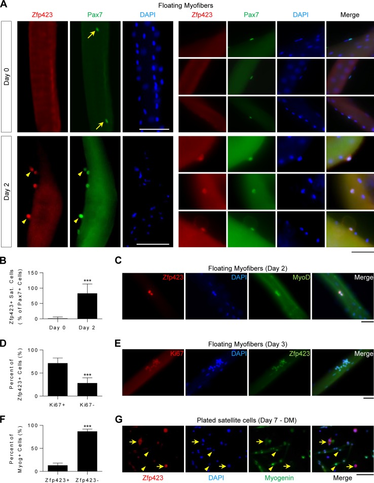
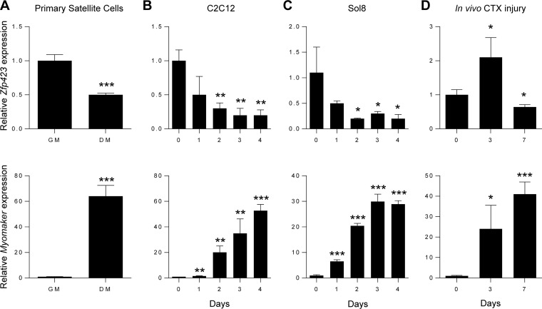
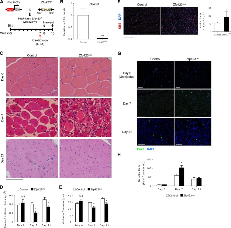
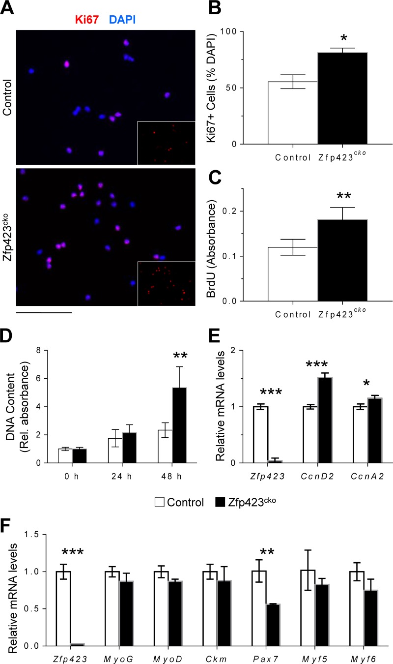
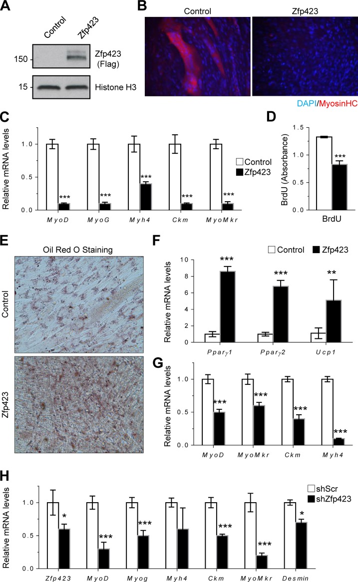
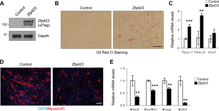
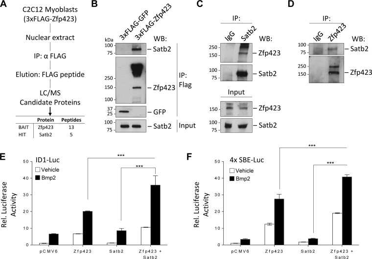
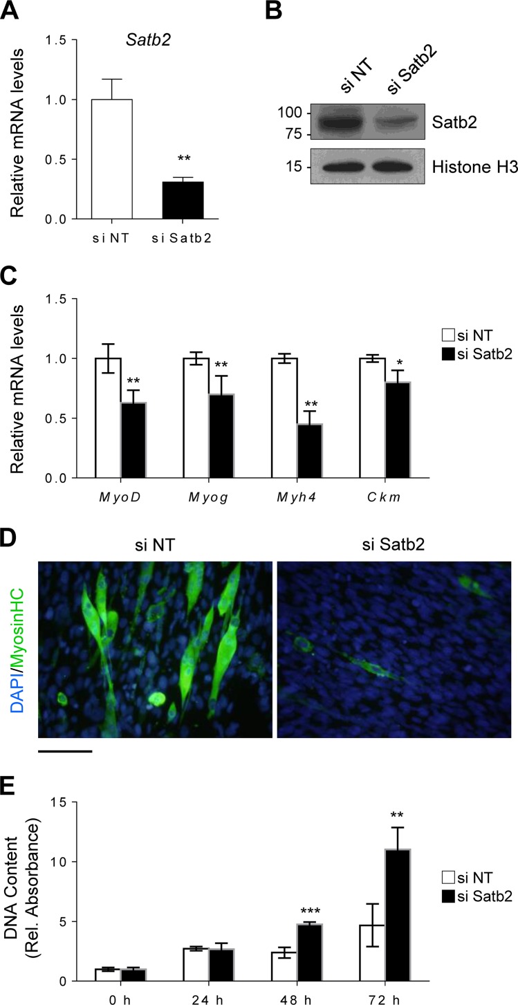
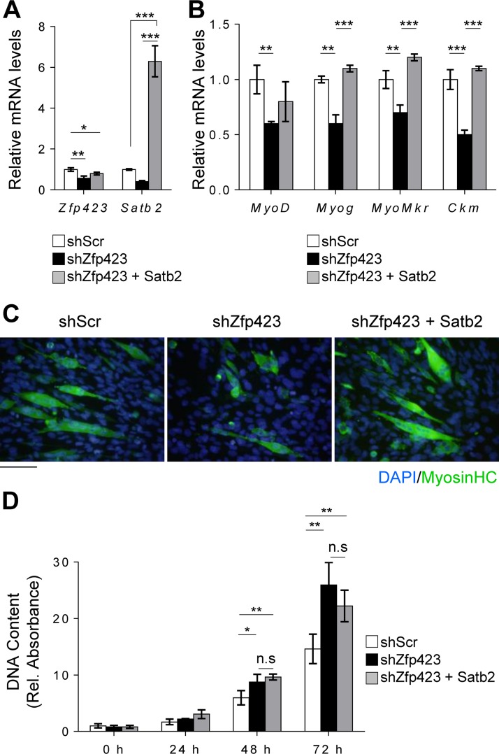
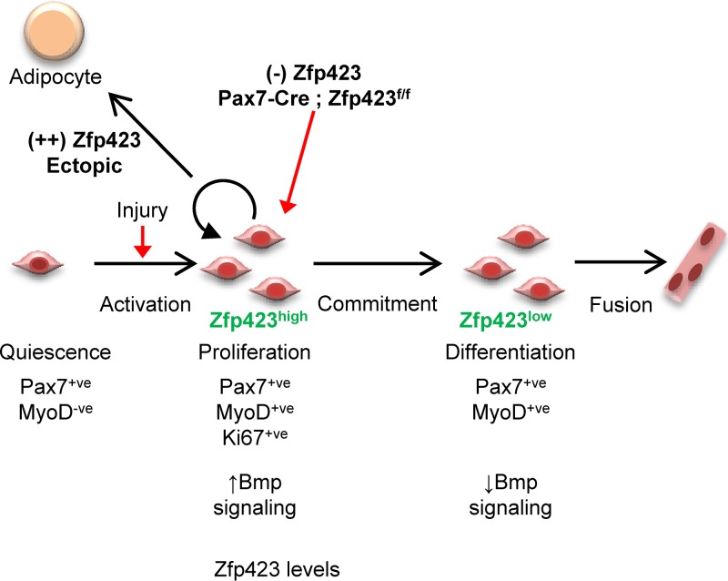
Satellite cells (SCs) are skeletal muscle stem cells that proliferate in response to injury and provide myogenic precursors for growth and repair. Zfp423 is a transcriptional cofactor expressed in multiple immature cell populations, such as neuronal precursors, mesenchymal stem cells, and preadipocytes, where it regulates lineage allocation, proliferation, and differentiation. Here, we show that Zfp423 regulates myogenic progression during muscle regeneration. Zfp423 is undetectable in quiescent SCs but becomes expressed during SC activation. After expansion, Zfp423 is gradually downregulated as committed SCs terminally differentiate. Mice with satellite-cell-specific Zfp423 deletion exhibit severely impaired muscle regeneration following injury, with aberrant SC expansion, defective cell cycle exit, and failure to transition efficiently from the proliferative stage toward commitment. Consistent with a cell-autonomous role of Zfp423, shRNA-mediated knockdown of Zfp423 in myoblasts inhibits differentiation. Surprisingly, forced expression of Zfp423 in myoblasts induces differentiation into adipocytes and arrests myogenesis. Affinity purification of Zfp423 in myoblasts identified Satb2 as a nuclear partner of Zfp423 that cooperatively enhances Zfp423 transcriptional activity, which in turn affects myoblast differentiation. In conclusion, by controlling SC expansion and proliferation, Zfp423 is essential for muscle regeneration. Tight regulation of Zfp423 expression is essential for normal progression of muscle progenitors from proliferation to differentiation.

Keywords: cell fate; myoblast; myogenesis; regeneration; satellite cells; skeletal muscle.


Figures

FIG 1
FIG 1
Zfp423 is expressed during satellite cell activation. (A to E) Freshly isolated myofibers, along with satellite cells in their niche, were immediately fixed (day 0) or suspension cultured in mitogen-rich activation medium for 2 to 3 days before fixation and immunostaining for Zfp423 and Pax7, MyoD, or Ki67. (A) Arrows indicate Zfp423-negative, Pax7-positive nuclei. Arrowheads indicate satellite cells doubly positive for both Zfp423 and Pax7. (B) Quantification of the number of Zfp423-positive satellite cells in the on days 0 and 2 of myofiber culture. (D) Quantification of the percentage of Zfp423-positive cells that express Ki67, as illustrated in panel E. (F and G) Satellite cells plated and differentiated for 7 days were stained for myogenin and Zfp423. The percentage of terminally differentiated (myogenin-positive) cells that express Zfp423 was quantified (F), and cells were imaged (G). (G) Arrows indicate Zfp423-positive, myogenin-positive cells, and arrowheads indicate Zfp423-positive, myogenin-negative cells. The data are presented as means ± the SD. ***, P < 0.001 (Student's t test). A total of >100 fibers were analyzed. Scale bars: 40 μm (A, C, and E) and 100 μm (G).
FIG 2
FIG 2
Zfp423 expression is downregulated during myogenic differentiation. (A) Primary satellite cells were plated and cultured in adherent culture conditions in growth medium (GM) or differentiation medium (DM) for 3 days, after which qRT-PCR analysis of Zfp423 and Myomaker expression was performed. (B) qRT-PCR of Zfp423 and Myomaker gene expression in undifferentiated proliferating C2C12 cells (day 0) and over the course of 4 days after the induction of myoblast differentiation. (C) qRT-PCR of Zfp423 and Myomaker expression in Sol8 myoblast cells during differentiation for the indicated number of days. (D) qRT-PCR showing Zfp423 and Myomaker expression during the course of muscle regeneration. TA muscles were injected with cardiotoxin (CTX) and harvested on the indicated days after injury for RNA. The data are presented as means ± the SD (n ≥ 3). *, P < 0.05; **, P < 0.01; ***, P < 0.001 (relative to GM or day 0; Student's t test).
FIG 3
FIG 3
Zfp423 is critical for muscle regeneration in vivo. (A) Schematic of CTX injury model. (B) Expression of Zfp423 in TA muscles of 7-week-old Pax7Cre; Zfp423f/f (Zfp423cko) mice. (C) H&E staining of TA muscles from control and Zfp423cko mice in uninjured conditions (day 0), as well as 7 and 21 days after injury. (D and E) Cross-sectional area (D) and minimal Feret’s diameter (E) of regenerating myofibers at the indicated time points following injury. (F) Ki67 immunostaining and quantification on control and Zfp423cko TA muscles at day 7 after injury. (G and H) Pax7 immunostaining (G) and quantification on control and Zfp423cko TA muscles (H) at days 0, 7, and 21 after injury. The data are presented as means ± the SD (n ≥ 3). n.s, not significant; *, P < 0.05; ***, P < 0.001 (Student's t test). Scale bars: 65 μm (C) and 100 μm (F and G).
FIG 4
FIG 4
Zfp423 impairs proliferative ability in satellite cells. (A to C) MACS-isolated wild-type and Zfp423cko satellite cells were immunostained and quantified for Ki67 and DAPI (A and B) or labeled with BrdU for 5 h for quantification of BrdU incorporation by anti-BrdU ELISA (C). (D) Wild-type and Zfp423cko satellite cells were plated and grown for 72 h, and proliferation was assessed by DNA quantification. (E) qRT-PCR of cell cycle regulators cyclin D2 and cyclin A2 in satellite cells grown as for panel D. (F) qRT-PCR of wild-type and Zfp423cko satellite cells cultured in differentiation medium (DM) for 3 days. The data are presented as means ± the SD (n ≥ 3). *, P < 0.05; **, P < 0.01; ***, P < 0.001 (Student's t test). Scale bar, 40 μm.
FIG 5
FIG 5
Forced expression of Zfp423 stimulates adipogenic differentiation in skeletal myoblasts. (A) Confirmation of Zfp423 overexpression by anti-FLAG Western blotting. (B) Sol8 cells stably expressing Zfp423 were induced to differentiate for 3 days, after which terminally differentiated myotubes were visualized by anti-MHC immunostaining. (C) qRT-PCR of myogenesis-selective genes in the differentiated cultures shown in panel B. (D) Sol8 cells stably expressing Zfp423 were labeled with BrdU for 5 h, and BrdU incorporation was quantified by anti-BrdU ELISA. (E and F) Sol8 cells stably expressing Zfp423 were cultured in adipogenic differentiation medium for 6 days, after which Oil Red O staining of lipid accumulation (E) or qRT-PCR analysis of adipocyte-specific genes (F) was performed. (G) qRT-PCR of myoblast-specific genes in the cultures described in panels D and E. (H) Sol8 cells expressing Zfp423 shRNA were cultured in myogenic differentiation medium for 3 days, after which myogenesis-selective genes were measured by qRT-PCR. The data are presented as means ± the SD (n ≥ 3). *, P < 0.05; **, P < 0.01; ***, P < 0.001 (Student's t test).
FIG 6
FIG 6
Zfp423 stimulates adipogenic differentiation in C2C12 myoblasts. (A) Confirmation of Zfp423 overexpression by anti-FLAG Western blotting. (B and C) C2C12 cells stably expressing Zfp423 were cultured in adipogenic differentiation medium for 6 days, after which Oil Red O staining of lipid accumulation (B) or qRT-PCR analysis of adipocyte-specific genes (C) was performed. (D and E) C2C12 cells stably expressing Zfp423 were cultured in myogenic differentiation medium for 3 days, after which terminally differentiated myotubes were visualized by anti-MHC immunostaining (D) and myogenesis-selective genes were analyzed by qRT-PCR (E). The data are presented as means ± the SD (n ≥ 3). **, P < 0.01; ***, P < 0.001 (Student's t test). Scale bars, 100 μm.
FIG 7
FIG 7
Zfp423 associates with Satb2 to cooperatively regulate transcription. (A) Schematic overview of the proteomics approach used to identify Zfp423 interacting proteins in C2C12 myoblast cells. FLAG-Zfp423 was purified from nuclear extracts with anti-FLAG antibody resin and analyzed by MS. See Table S1 in the supplemental material for a complete list of interactants. (B) Immunoprecipitation analysis of FLAG-Zfp423 with endogenous Satb2. C2C12 cells stably expressing FLAG-Zfp423 or FLAG-GFP were lysed and immunoprecipitated (IP) with anti-FLAG antibody and blotted (WB) with anti-FLAG antibody or anti-Satb2 antibody. (C and D) Confirmation of endogenous Zfp423 interaction with endogenous Satb2. Zfp423 or Satb2 was immunoprecipitated from untransfected C2C12 cells with anti-Zfp423 or anti-Satb2 antibody and immunoblotted with the indicated antibody. Naive IgG was used as a negative-control antibody. (E) Zfp423 works in concert with Satb2. The transcriptional activity of a Bmp-responsive Id1 promoter luciferase (ID1-Luc) construct in the presence of the indicated expression vectors for Zfp423 or Satb2 was assessed. (F) Luciferase activity of a BMP-responsive construct containing four tandem repeats of the SMAD binding elements (SBE-Luc) in cells transfected with the indicated expression vectors. All luciferase activity was measured 16 h after treatment with 100 ng/ml BMP2. relative to the empty-vector control unless otherwise indicated. The data are presented as means ± the SD (n ≥ 3). ***, P < 0.001 (Student's t test).
FIG 8
FIG 8
Satb2 regulates myoblast differentiation and proliferation. (A and B) Satb2 mRNA (A) and protein (B) were efficiently depleted by siRNA-mediated knockdown in Sol8 cells. Nontargeting siRNA (si NT) was used as a control. (C and D) Satb2 knockdown cells were induced to differentiate for 3 days, after which myogenesis-selective genes were analyzed by qRT-PCR (C) and terminally differentiated myotubes were visualized by anti-MHC immunostaining (D). (E) Satb2 knockdown cells and nontargeting siRNA control cells were seeded at a low density and cultured for 72 h, during which proliferation was monitored. The data are presented as means ± the SD (n ≥ 3). *, P < 0.05; **, P < 0.01; ***, P < 0.001 (Student's t test). Scale bar, 100 μm.
FIG 9
FIG 9
Functional interaction between Zfp423 and Satb2 in myogenesis. Sol8 cells stably expressing Zfp423 shRNA (shZfp423) or scrambled shRNA (shScr) were transfected with control (empty vector) or Satb2 expression vectors. (A) After selection, qRT-PCR confirmed the overexpression of Satb2. (B and C) Cells were then differentiated for 3 days in myogenic differentiation medium, and the expression of myogenesis-selective genes was quantified by qRT-PCR (B) and differentiated myotubes were visualized by anti-MHC immunostaining (C). (D) DNA content as a measure of cell proliferation. The data are presented as means ± the SD (n ≥ 3). *, P < 0.05; **, P < 0.01; ***, P < 0.001 (ANOVA with a Fisher least-significant-difference post hoc test). Scale bar, 100 μm.
FIG 10
FIG 10
Model for the role of Zfp423 in adult muscle regeneration. During adult muscle regeneration, quiescent Pax7+ satellite cells do not express Zfp423, but activated cells begin to express Zfp423. After extensive proliferation, cells committed to myogenic differentiation downregulate Zfp423 and fuse to form myotubes. Loss of Zfp423 results in a delay of progression from the proliferative state to the myogenic differentiation state. Thus, Zfp423 controls the proliferation of adult satellite cells to affect muscle regeneration and myogenesis.

References

    1. Pittenger MF, Mackay AM, Beck SC, Jaiswal RK, Douglas R, Mosca JD, Moorman MA, Simonetti DW, Craig S, Marshak DR. 1999. Multilineage potential of adult human mesenchymal stem cells. Science 284:143–147. doi:10.1126/science.284.5411.143. - DOI - PubMed
    1. Fazeli PK, Horowitz MC, MacDougald OA, Scheller EL, Rodeheffer MS, Rosen CJ, Klibanski A. 2013. Marrow fat and bone—new perspectives. J Clin Endocrinol Metab 98:935–945. doi:10.1210/jc.2012-3634. - DOI - PMC - PubMed
    1. Kaplan FS, Shore EM. 2000. Progressive osseous heteroplasia. J Bone Miner Res 15:2084–2094. doi:10.1359/jbmr.2000.15.11.2084. - DOI - PubMed
    1. Wosczyna MN, Biswas AA, Cogswell CA, Goldhamer DJ. 2012. Multipotent progenitors resident in the skeletal muscle interstitium exhibit robust BMP-dependent osteogenic activity and mediate heterotopic ossification. J Bone Miner Res 27:1004–1017. doi:10.1002/jbmr.1562. - DOI - PMC - PubMed
    1. Seale P, Bjork B, Yang W, Kajimura S, Chin S, Kuang S, Scime A, Devarakonda S, Conroe HM, Erdjument-Bromage H, Tempst P, Rudnicki MA, Beier DR, Spiegelman BM. 2008. PRDM16 controls a brown fat/skeletal muscle switch. Nature 454:961–967. doi:10.1038/nature07182. - DOI - PMC - PubMed

Publication types

MeSH terms

LinkOut - more resources