. 2019 Jan 8:12:505.
doi: 10.3389/fncel.2018.00505. eCollection 2018.

Herpes Simplex Virus Type 1 Enhances Expression of the Synaptic Protein Arc for Its Own Benefit

Affiliations

Herpes Simplex Virus Type 1 Enhances Expression of the Synaptic Protein Arc for Its Own Benefit

Francisca Acuña-Hinrichsen et al. Front Cell Neurosci. .

Abstract

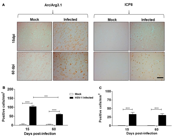
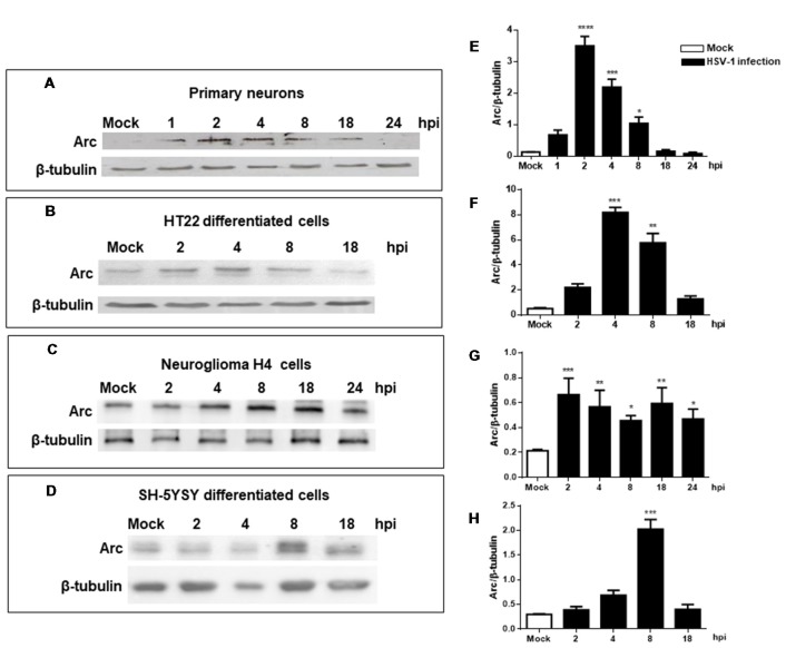
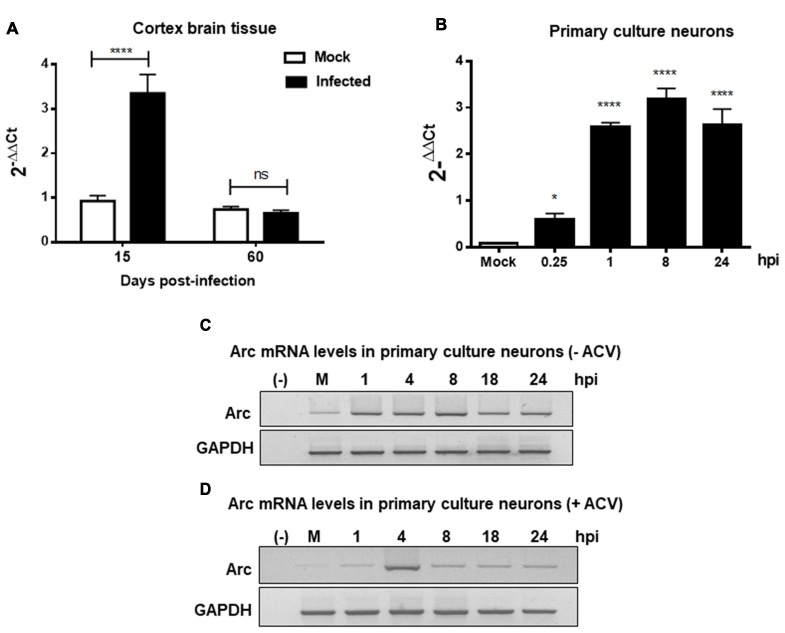
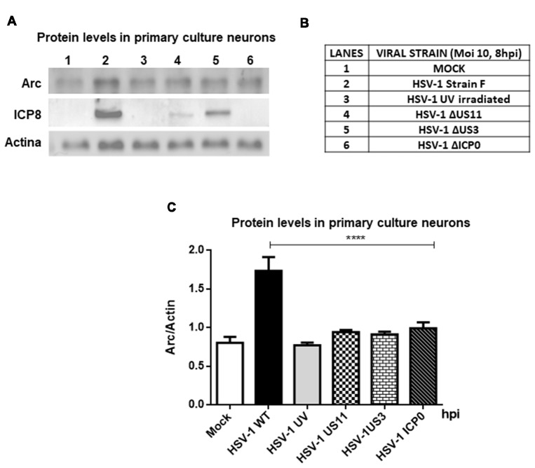
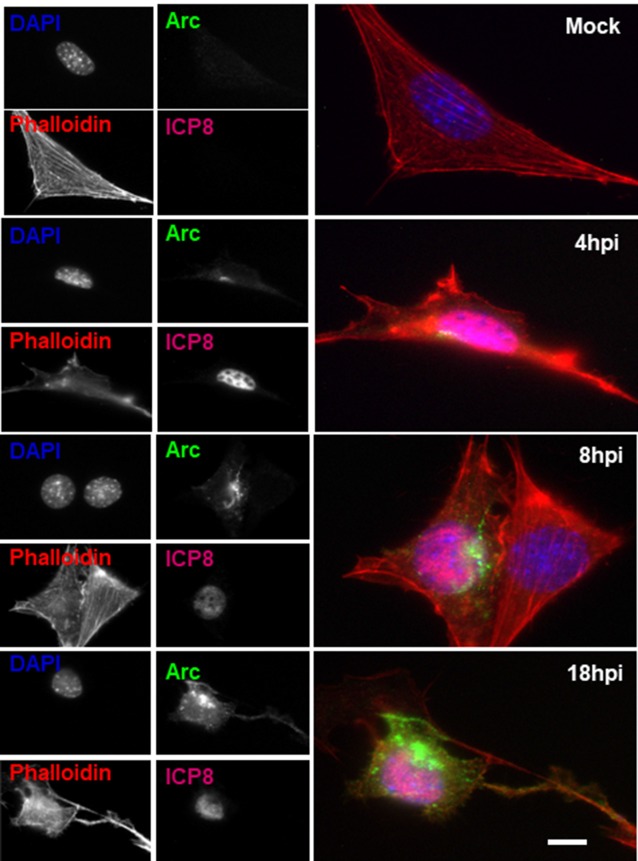
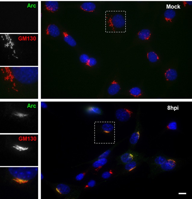
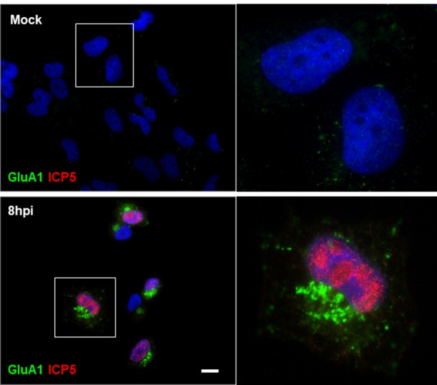
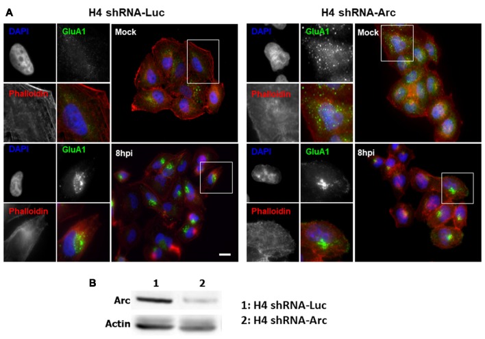
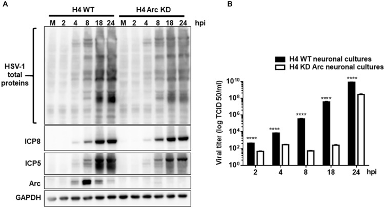
Herpes simplex virus type 1 (HSV-1) is a neurotropic virus able to reach the central nervous system (CNS) after primary infection in oronasal mucosa. HSV-1 establishes latency inside neurons due the repression of its gene expression process, which is related to periodic reactivations in response to cellular stress conditions, constituting a risk factor for neurodegenerative diseases such as Alzheimer's disease (AD). The immediate-early gene Arc plays an essential role in neuronal morphology, synaptic plasticity and memory formation. Arc acts as a hub protein, interacting with components of the endocytic machinery required for AMPA receptor (AMPAR) recycling as well as with proteins of the post-synaptic density and actin cytoskeleton. However, to date, no studies have evaluated whether persistent neurotropic HSV-1 infection modulates the expression or function of Arc protein in brain tissue. Here, we report that neuronal in vivo and in vitro infection of HSV-1 significantly increases Arc protein levels, showing a robust perinuclear distribution in neuronal cell lines, a process that is dependent on an active HSV-1 replication cycle. Finally, we found that silencing Arc protein caused a decrease in HSV-1 proteins and viral progeny, suggesting that Arc is involved in the lifecycle of HSV-1. Our studies strongly suggest that pathogenicity of HSV-1 neuronal reactivations in humans could be mediated in part by Arc neuronal upregulation and its potential role in endocytic trafficking and AMPA-neuronal function impairment. Further studies are necessary to define whether this phenomenon could have repercussions in cognition and learning processes in infected individuals.

Keywords: Alzheimer’s disease; Arc; HSV-1; neurodegeneration; neuronal dysfunction; neuronal infection; neurotropic virus.


Figures

Figure 1
Figure 1
Herpes simplex virus type 1 (HSV-1)-infected mice present elevated levels of Arc protein. (A) Cortexes from mock and HSV-1 infected mice (six animals per time and condition) were stained at 15, 60 days post-infection (dpi) post-infection with antibodies to Arc and to ICP8 (three slices per animal). Magnification 100×, scale bar represents 100 nm. (B,C) Graphics show the number of Arc/ICP8 positive cells/mm2 (n = 3 sections per times). ****p < 0.0001 and ***p < 0.001 [analyzed by Two-Way analyses of variance (ANOVA)]. Statistical analyses between mock times to Arc and ICP8 were not significant. Also were not significant to ICP8 in infected times at 15 dpi compared to 60 dpi.
Figure 2
Figure 2
Increased levels of Arc protein during in vitro neuronal HSV-1 infection. Immunoblot analyses showing Arc and β-tubulin proteins, in either non-infected (Mock or M) or HSV1-infected brain cells multiplicity of infection (MOI = 10). (A) Mouse primary culture neurons. (B) Differentiated HT22 cells. (C) Neuroglioma H4 cells. (D) Differentiated SH-SY5Y cells. Infections were performed at 1, 2, 4, 8, 18 and 24 hours post-infection (hpi). (E–H) After gel scanning and densitometry quantitation, the results were expressed as the ratio of Arc protein levels respect to total tubulin. The blots are representative of three independent experiments. ****p < 0.0001; ***p < 0.001; **p < 0.01; *p < 0.05 (analyzed by One-Way ANOVA, followed by Tukey’s multiple comparisons Test).
Figure 3
Figure 3
HSV-1 neuronal infection and replication triggers upregulation of Arc mRNA levels. Arc mRNA levels were analyzed by RT-qPCR in either (A) Brain cortex of mock and HSV-1 infected mice at 15 or 60 dpi or (B) mouse primary culture neurons infected during 24 h with HSV-1 and mock conditions. Relative Arc mRNA expression levels were controlled in respect to the levels of the housekeeping gene PPIA. After gel scanning and densitometry quantitation, the results were expressed as the ratio of Arc mRNA in respect to PPIA mRNA levels. Experiments were performed in triplicate. ****p > 0.0001 and *p > 0.05; ns: non-significant (using Two-Way ANOVA analyze in graphic A, and One-Way ANOVA analyze in graphic B). Conventional RT-PCR in Mock- and HSV-1-infected (MOI = 10) mouse primary culture neurons during 1, 4, 8, 18 and 24 hpi without (C) or with (D) acyclovir (ACV) treatment added during the time course of infection.
Figure 4
Figure 4
Viral replication is mandatory to induce an increase in Arc protein levels. (A) Immunoblot analyses showing Arc, ICP8, Actin and GAPDH proteins, in either mock or HSV-1-infected neurons (lanes 1–6) with a MOI of 10 and at 8 hpi. (B) Table indicating the strains tested including Strain F (wild-type HSV-1) and different replicative-defective HSV-1 mutants and UV-irradiated (inactivated) HSV-1. (C) After gel scanning and densitometry quantitation, the results were expressed as the ratio of Arc protein levels in respect to total Actin. The results shown are representative of three independent experiments. ****p > 0.0001 (analyzed by one-way ANOVA, followed by Tukey’s multiple comparisons test).
Figure 5
Figure 5
Altered Arc protein distribution during HSV-1 neuronal infection. Untreated (mock) or HSV-1-infected HT22 cells (MOI = 10) during 4, 8 or 18 hpi were stained with specific antibodies to Arc or to ICP8 followed by secondary antibodies coupled to Alexa Fluor 488 and 647 dyes, respectively. Phalloidin staining was coupled with Alexa Fluor 594 dye and Nuclei stained with DAPI. Scale bar corresponds to 10 μm. Magnification 630×. The results shown are representative of three independent experiments.
Figure 6
Figure 6
HSV-1 infection induced colocalization of Arc protein with the Golgi marker GM130 in HT22 cells. Untreated (mock) or HSV-1-infected (MOI = 10) HT22 differentiated cells during 8 hpi were stained with antibodies to Arc and the Golgi marker GM130 followed by secondary antibodies coupled to Alexa Fluor 488 and 594 dyes, respectively. Nuclei were stained with DAPI. Scale bar corresponds to 10 μm. Magnification 630×. The box inserted shows a zoom capture of the indicated area. The results shown are representative of three independent experiments.
Figure 7
Figure 7
Redistribution of the AMPA receptor (AMPAR) GluA1 subunit during HSV-1 infection in H4 cells. Untreated (mock) or HSV-1-infected (MOI = 10) H4 cells during 8 hpi were stained with antibodies to GluA1 subunit and to ICP5 followed by secondary antibodies coupled to Alexa Fluor 488 and 594 dyes, respectively. Nuclei were stained with DAPI. Bar corresponds to 10 μm. Magnification 630×. The boxes inserted show a zoom capture of the indicated areas. The results shown are representative of three independent experiments.
Figure 8
Figure 8
Redistribution of AMPAR GluA1 subunit during HSV-1 infection is independent of Arc expression. (A) H4 cells stably expressing either the shRNA to luciferase (H4 shRNA-Luc) used as a control or the shRNA to Arc (H4 shRNA-Arc) to achieve silencing of Arc knockdown (KD) were infected with HSV-1 (MOI = 10) during 8 hpi. Then, GluA1 subunit and ICP8 were immunodetected with specific antibodies followed of secondary antibodies couple to Alexa Fluor 488 and 647, respectively. Phalloidin staining was coupled with Alexa Fluor 594 dye and Nuclei stained with DAPI. Scale bar corresponds to 10 μm. Magnification 630×. The boxes inserted show a zoom capture of the indicated areas. (B) Total protein extracts of H4 shRNA-Luc and H4 shRNA-Arc were analyzed by immunoblot with antibodies to the proteins Arc and GADPH. The results shown are representative of three independent experiments.
Figure 9
Figure 9
Arc enhances HSV-1 viral progeny in vitro. (A) H4 cells stably expressing either the shRNA to luciferase (H4 shRNA-Luc) used as a control or the shRNA to Arc (H4 shRNA-Arc) to achieve silencing of Arc KD were infected with HSV-1 (MOI = 10) during 2, 4, 8, 12, 18 y 24 hpi. First, infected cells were harvested and the production of infectious viral progeny in the supernatants measured in each time point by a standard TCID50 assay. Second, total protein extracts were analyzed by immunoblot with antibodies to the proteins ICP8, ICP5, Arc and GADPH. (B) Graphic shows the viral titer of HSV-1 in the supernatants from H4 shRNA-Luc (black bars) or H4 shRNA-Arc (white bars). The results shown are representative of three independent experiments (****p > 0.0001, analyzed by two-way ANOVA).

Similar articles

Cited by

References

    1. Ando Y., Kitayama H., Kawaguchi Y., Koyanagi Y. (2008). Primary target cells of herpes simplex virus type 1 in the hippocampus. Microbes Infect. 10, 1514–1523. 10.1016/j.micinf.2008.09.005 - DOI - PubMed
    1. Ashley J., Cordy B., Lucia D., Fradkin L. G., Budnik V., Thomson T. (2018). Retrovirus-like gag protein arc1 binds RNA and traffics across synaptic boutons. Cell 172, 262.e11–274.e11. 10.1016/j.cell.2017.12.022 - DOI - PMC - PubMed
    1. Bearer E. L. (2012). HSV, axonal transport and Alzheimer’s disease: in vitro and in vivo evidence for causal relationships. Future Virol. 7, 885–899. 10.2217/fvl.12.81 - DOI - PMC - PubMed
    1. Bliss T. V., Collingridge G. L. (1993). A synaptic model of memory: long-term potentiation in the hippocampus. Nature 361, 31–39. 10.1038/361031a0 - DOI - PubMed
    1. Bramham C. R. (2007). Control of synaptic consolidation in the dentate gyrus: mechanisms, functions, and therapeutic implications. Prog. Brain Res. 163, 453–471. 10.1016/s0079-6123(07)63025-8 - DOI - PubMed

LinkOut - more resources