

Review
doi: 10.4274/tjod.88614.
Epub 2019 Jan 9.
Basic clinical retroperitoneal anatomy for pelvic surgeons
Affiliations
- PMID: 30693143
- PMCID: PMC6334244
- DOI: 10.4274/tjod.88614
Item in Clipboard
Review
Basic clinical retroperitoneal anatomy for pelvic surgeons
Turk J Obstet Gynecol.
2018 Dec.
Abstract
Basic anatomical knowledge should be improved during residency period with clinical practice. Especially pelvic surgeons; obstetricians, gynecologists, gynecological oncologists, urologists and general surgeons must have an advanced level practise of retroperitoneal anatomy to gain surgical skills. Retroperitoneal topographic anatomy, retroperitoneal vasculature, ureteric dissection and pelvic avascular spaces are the precise points during pelvic surgery.
Keywords: Surgery; anatomy; gynecology; hypogastric; ureter.
Conflict of interest statement
Conflict of Interest: No conflict of interest was declared by the authors.
Figures
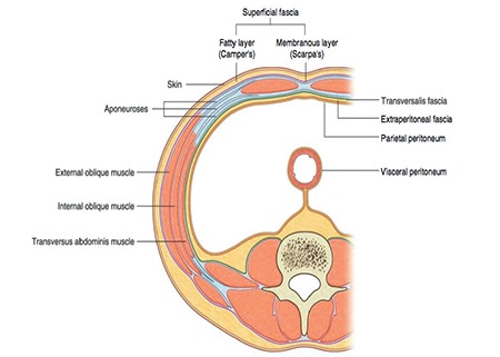
Transverse section of the anterior abdominal wall; the extraperitoneal fascia with fatty tissue under the parietal peritoneum lies on the posterior abdominal wall called the retroperitoneum (Gray’s Anatomy for Students, 3rd Edition, Churchill Livingstone/Elsevier, 2015)(1)
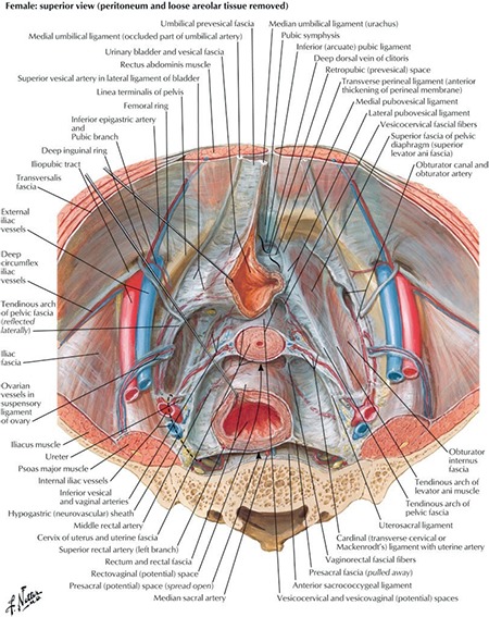
Pelvic viscera and retroperitoneum (Atlas of Human Anatomy, 6th Edition, Saunders/Elsevier, 2014)(2)
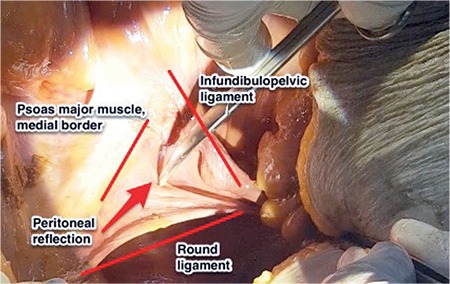
Demonstration to enter the retroperitoneum between the round ligament and infundibulopelvic ligament (lateral parietal peritoneum), right pelvic side wall (cadaveric dissection)
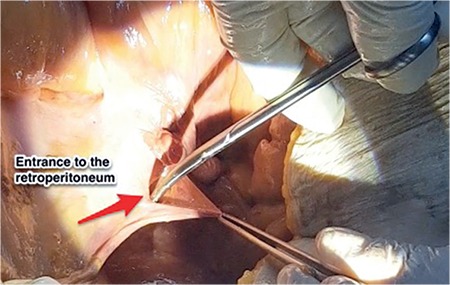
Demonstration of opening retroperitoneum, right pelvic side wall (cadaveric dissection)
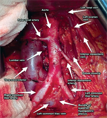
Paraaortic region, aorta and inferior vena cava after paraaortic lymphadenectomy (surgical archieve)
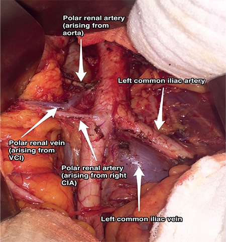
Polar renal artery arising from the right common iliac artery and also abdominal aorta (surgical archive) VCI: Vena cava inferior, CIA: Common iliac artery
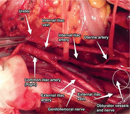
Uterine artery, right pelvic side wall (surgical archive)
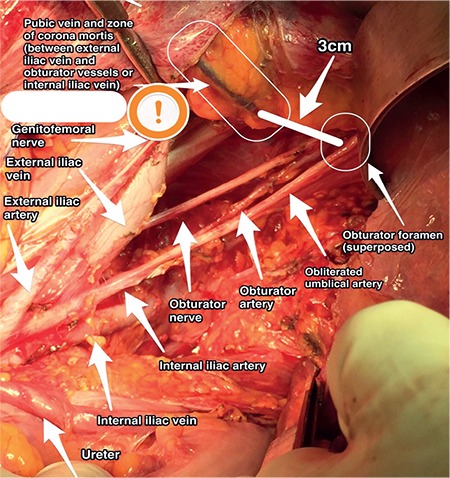
Pubic vein, left pelvic side wall (surgical archive)
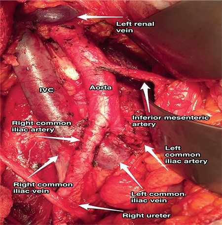
Left and right common iliac veins and arteries (surgical archive) IVC: Inferior vena cava
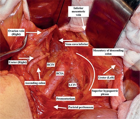
Right ureter below the right ovarian vein medial to the ascending colon and lateral to inferior vena cava, and left ureter underneath the mesentery of descending colon, medial/ parallel to the inferior mesenteric vein and lateral to aorta/ superior hypogastric plexus (surgical archive) RCIV: Right common iliac vein, RCIA: Right common iliac artery, LCIV: Left common iliac vein
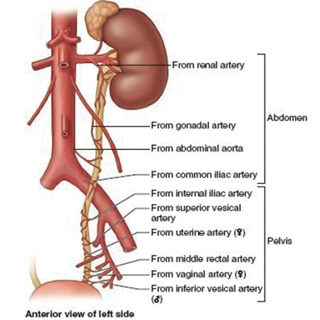
Vascularization of ureter from the kidney to the bladder (left side), while dissecting the ureter traction should be applied towards the side of blood vessels (Moore Clinically Oriented Anatomy, 7th Edition, Wolters Kluwer/Lippincott Williams & Wilkins, 2013)(6)
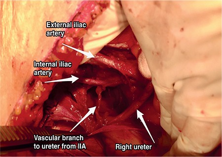
Vascular branch to ureter from internal iliac artery, right pelvic side wall, in the pelvis the most important vascular supply of the ureter is the branch from the internal iliac artery (surgical archive) IIA: Internal iliac artery
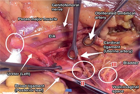
Sites of ureter injury, left pelvic side wall: Zone I, during infundibulopelvic ligament ligation just below the level of pelvic inlet; zone II, during uterine artery ligation (ureter crosses the cardinal ligament-uterine artery complex); zone III, during vaginal excision (ureter is anterolateral to the anterior vagina before entering the bladder-trigone) (cadaveric dissection) EIA: External iliac artery
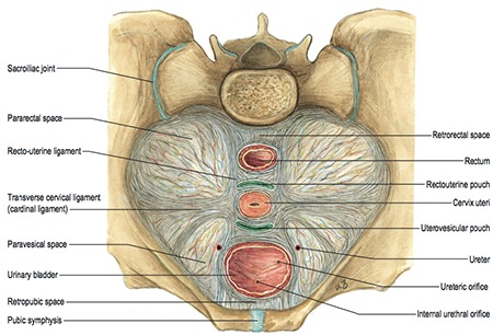
Avascular spaces and supporting ligaments in the pelvis (Sobotta Atlas of Human Anatomy, 15th Edition, Elsevier, Urban&Fischer. Copyright 2013/Gray’s Anatomy, The Anatomical Basis of Clinical Practice, 41th edition, Elsevier, 2016)(7)
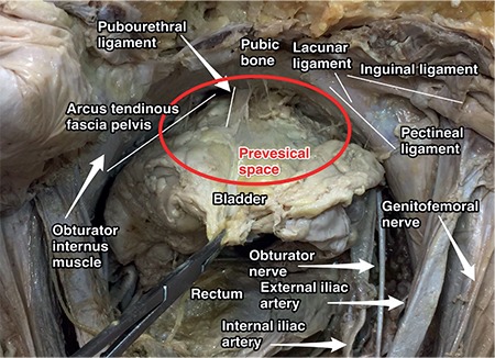
Prevesical space and contents (cadaveric dissection)
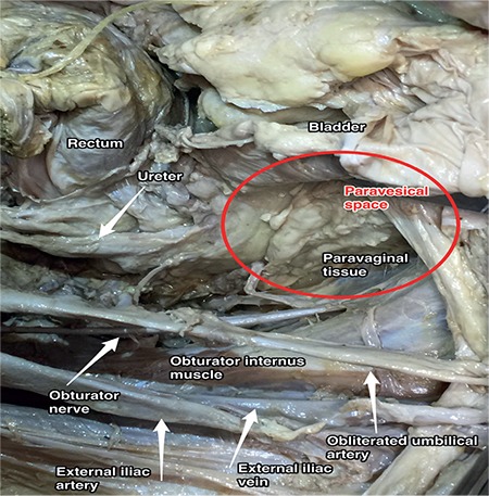
Paravesical space, right pelvic side wall (cadaveric dissection)
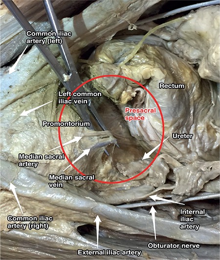
Presacral space (cadaveric dissection)
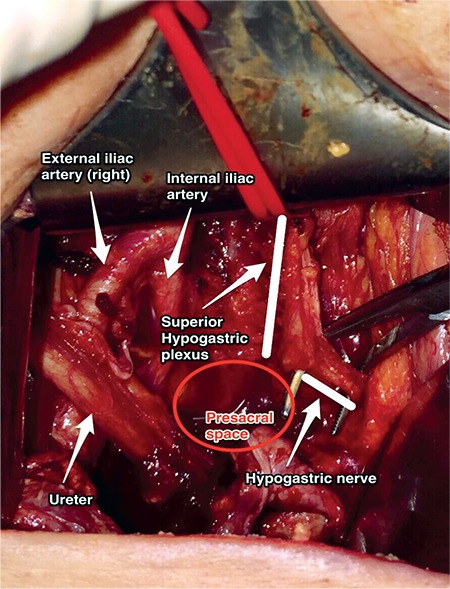
Presacral space and superior hypogastric plexus (surgical archive)
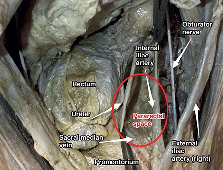
Pararectal space, right pelvic side wall (cadaveric dissection)
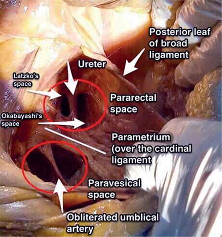
Right pelvic side wall; the paravesical space, anterior to the cardinal ligament is divided into two parts by the obliterated umbilical artery and the pararectal space, posterior to the cardinal ligament is divided into two parts by the ureter, the lateral part is called Latzko’s space and the medial part is called Okabayashi’s space (cadaveric dissection)
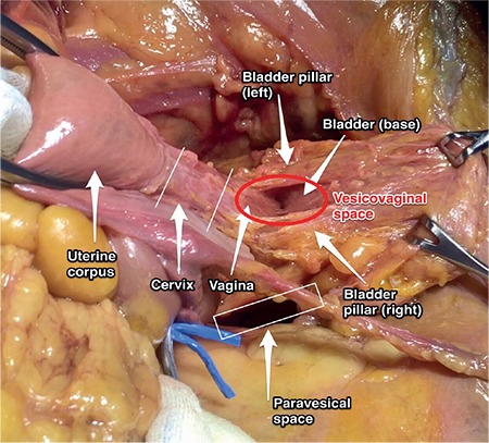
Vesicovaginal space (cadaveric dissection)
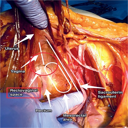
Rectovaginal space (cadaveric dissection)
References
-
- Richard Drake, Wayne Vogl and Adam W M. Mitchell, Gray’s Anatomy for Students, 3rd Edition. Churchill Livingstone/Elsevier. 2015.
-
- Frank Netter. Atlas of Human Anatomy, 6th Edition. Saunders/ Elsevier. 2014.
-
- Murat Öz, Salim Erkaya, Bülent Özdal, Mehmet Mutlu Meydanlı, İlker Selçuk, Tayfun Güngör. Retroperitoneal vasküler varyasyonlar ve jinekolojik onkoloji cerrahisinde önemi. Türk Jinekolojik Onkoloji Dergisi. 2014:123–8.
-
- Tuğba Tekelioğlu, Hasan Aykut Tuncer, Eda Adeviye Şahin, İlker Selçuk. Pelvisin Vasküler Anatomisi. In: Ali Ayhan, Hüsnü Çelik, Polat Dursun, editor. Jinekolog Onkolog Bakış Açısıyla; Postpartum Kanama. Ankara/Turkey. Güneş Tıp Kitabevleri. 2017.
Publication types
LinkOut - more resources
Full Text Sources