

Prenatal detection of Peters plus-like syndrome
- PMID: 30693145
- PMCID: PMC6334245
- DOI: 10.4274/tjod.45649
Prenatal detection of Peters plus-like syndrome
Abstract
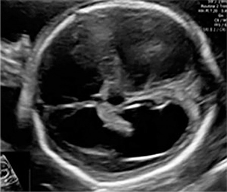
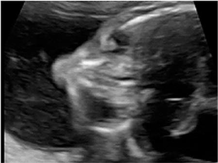
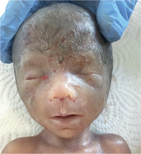
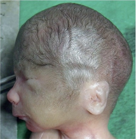
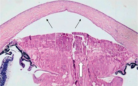
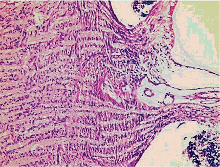
Peters plus syndrome is a rare congenital disorder that includes ocular anterior segment defects of the classic Peter's anomaly, and is mostly associated with craniofacial and skeletal defects. A 21-week fetus was referred for further evaluation due to a suspicion of fetal hydrocephalus. An ultrasound examination revealed hyperechogenic lenses, microphthalmia, hypotelorism, retrognathia, mild ventriculomegaly, absence of the cavum septum pellucidum, and short stature. Amniocentesis and further microarray analysis revealed normal chromosomal copy numbers including the gene B3GALTL. In utero mort fetalis occurred at the 23rd gestational week. Ultrasound and fetal autopsy findings were suggestive of Peters plus syndrome, but the absence of the B3GALTL gene mutation made the diagnosis Peters plus-like syndrome. Obstetricians should consider Peters plus-like syndrome with prenatal detection of ocular anomalies along with craniofacial and skeletal anomalies with the absence of B3GALTL gene mutation.
Keywords: B3GALTL gene; Peters anomaly; Peters plus syndrome; congenital cataract; prenatal diagnosis.
Conflict of interest statement
Conflict of Interest: The authors declare no conflict of interest.
Figures
References
-
- Peters A. Ueber angeborene Defektbildung der Descemetschen Membran. Klin Monatsbl Augenheilkd. 1906;44:27–40.
-
- Maillette de Buy Wenniger-Prick LJ, Hennekam RC. The Peters’ plus syndrome: a review. Ann Genet. 2002;45:97–103. - PubMed
-
- Boog G, Le Vaillant C, Joubert M. Prenatal sonographic findings in Peters-plus syndrome. Ultrasound Obstet Gynecol. 2005;25:602–6. - PubMed
-
- Schoner K, Kohlhase J, Müller AM, Schramm T, Plassmann M, Schmitz R, et al. Hydrocephalus, agenesis of the corpus callosum, and cleft lip/palate represent frequent associations in fetuses with Peters’ plus syndrome and B3GALTL mutations. Fetal PPS phenotypes, expanded by Dandy Walker cyst and encephalocele. Prenat Diagn. 2013;33:75–80. - PubMed
Publication types
LinkOut - more resources
Full Text Sources
Miscellaneous