. 2018 Nov 19;6(2):1800361.
doi: 10.1002/advs.201800361. eCollection 2019 Jan 23.

Nanoscale Coatings for Ultralow Dose BMP-2-Driven Regeneration of Critical-Sized Bone Defects

Affiliations

Nanoscale Coatings for Ultralow Dose BMP-2-Driven Regeneration of Critical-Sized Bone Defects

Zhe A Cheng et al. Adv Sci (Weinh). .

Abstract

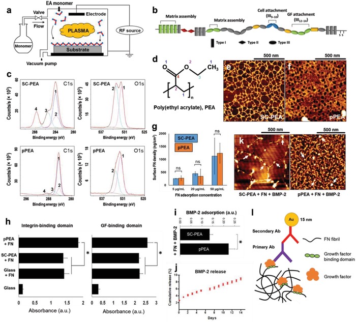
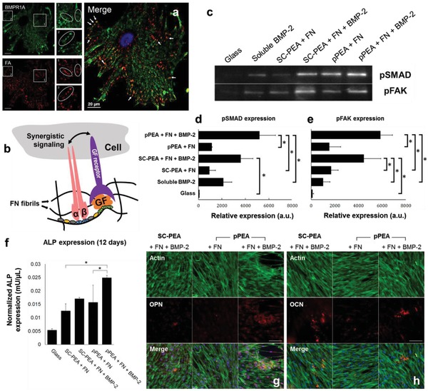
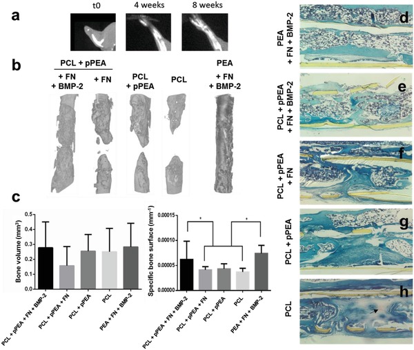
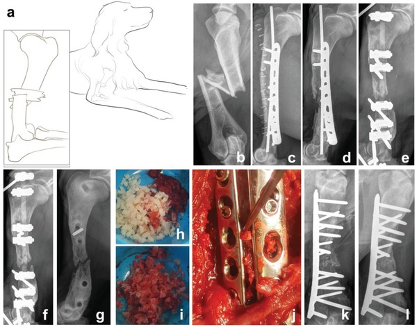
While new biomaterials for regenerative therapies are being reported in the literature, clinical translation is slow. Some existing regenerative approaches rely on high doses of growth factors, such as bone morphogenetic protein-2 (BMP-2) in bone regeneration, which can cause serious side effects. An ultralow-dose growth factor technology is described yielding high bioactivity based on a simple polymer, poly(ethyl acrylate) (PEA), and mechanisms to drive stem cell differentiation and bone regeneration in a critical-sized murine defect model with translation to a clinical veterinary setting are reported. This material-based technology triggers spontaneous fibronectin organization and stimulates growth factor signalling, enabling synergistic integrin and BMP-2 receptor activation in mesenchymal stem cells. To translate this technology, plasma-polymerized PEA is used on 2D and 3D substrates to enhance cell signalling in vitro, showing the complete healing of a critical-sized bone injury in mice in vivo. Efficacy is demonstrated in a Münsterländer dog with a nonhealing humerus fracture, establishing the clinical translation of advanced ultralow-dose growth factor treatment.

Keywords: biomaterials; bone regeneration; growth factor delivery; stem cell differentiation.


Figures

Figure 1
Figure 1
Physicochemical characterisation of plasma PEA coatings, FN, and BMP‐2 adsorption on pPEA. a) Schematic representation of a custom‐made plasma polymerization chamber. b) FN structure, showing its three domain types (I, II, III) and functions. Domain III region (III9–10) contains the RGD (Arg‐Gly‐Asp) sequence that facilitates cell adhesion via integrin binding, and region III12–14 binds various GFs, including BMP‐2. c) XPS characterization of SC‐PEA and pPEA. High‐resolution C1s and O1s spectra are shown with fitted components in colored dotted lines. d) Chemical structure of PEA, with labelled carbon and oxygen atoms corresponding to components in panel (c). AFM phase images of FN adsorbed for 10 min on e) SC‐PEA and f) pPEA. Thin fibrillar networks were observed on SC‐PEA, whereas thick, dense networks were observed on pPEA. g) Surface density of FN adsorbed at different concentrations onto SC‐PEA and pPEA for 1 h. h) Relative exposure of integrin‐binding and GF‐binding domains on FN adsorbed on different surfaces, measured using ELISA. i) Relative adsorption of BMP‐2 on FN‐coated surfaces, measured using ELISA. j) Cumulative BMP‐2 release from surfaces coated with pPEA, FN, and BMP‐2 during 2 weeks. k) AFM height images of BMP‐2 labelled with gold nanoparticles on SC‐PEA and pPEA coated with FN. White arrows indicate gold nanoparticles showing BMP‐2 distribution. l) Schematic representation of immunogold assay for GF detection. All data are presented as mean ± SD, n = 3, one‐way ANOVA with Tukey's test for multiple comparisons. *p < 0.05, ns = not statistically significant.
Figure 2
Figure 2
hMSC signalling and differentiation. a) Colocalization assay of BMP receptor 1A (BMPR1A, green) and FAs (red). White arrows on the merged image show areas of colocalization in yellow. Scale bar = 20 µm. b) A schematic representation showing that synergistic signalling between integrin and GF receptors can occur when the integrin‐binding (III9–10) and GF‐binding (III12–14) domains of FN are in close proximity. c) Western blotting of pSMAD 1/5/9 and pFAK, expressed by hMSCs after 1 h in culture on SC‐PEA and pPEA, with and without FN and BMP‐2. Quantified blots to show relative expressions of d) pSMAD and e) pFAK, both normalized using total protein amount, from hMSCs after 1 h in culture on SC‐PEA and pPEA, with and without FN and BMP‐2. hMSCs cultured with soluble BMP‐2 and on glass alone were used as controls. Data are presented as mean ± SD, n = 3, one‐way ANOVA with Tukey's test for multiple comparisons. *p < 0.05. f) Normalized ALP expression in hMSCs after 12 d in culture on SC‐PEA and pPEA surfaces, with and without FN and BMP‐2, from a fluorescent ALP assay. Data are presented as mean ± SD, n = 3, one‐way ANOVA with Tukey's test for multiple comparisons. *p < 0.05. Immunofluorescent labelling of g) OPN and h) OCN in hMSCs cultured on SC‐PEA and pPEA, with and without FN and BMP‐2, for 21 d. Phalloidin stains actin cytoskeleton in green and DAPI stains nuclei in blue. Scale bar = 50 µm.
Figure 3
Figure 3
Bone regeneration in a murine model of a critical‐sized radial bone defect with low doses of BMP‐2. a) X‐ray images at 0, 4, and 8 weeks after surgery. b) 3D reconstructions from the µCT images showing the radius in the area of the defect, 8 weeks after introduction of the PCL‐pPEA implant (with or without FN and BMP‐2). c) Quantification of the volume and specific surface of new bone. Data are presented as mean ± SD, minimum n = 3. Two‐tailed t‐test was used to analyze data. *p < 0.1. d–h) Hematoxylin‐Safranin O‐fast green staining of histological sections in the area of the defect. The tissue is organized in structures resembling bone marrow (rounded white structures in panels (d) and (e)) versus fibroblast‐like morphology (extended and aligned) in the center of the defect in panels (f)–(h). Arrow points to red staining that indicates cartilage formation in panel (h).
Figure 4
Figure 4
Humeral fracture healing in a dog treated with bone chips coated with pPEA, FN, and BMP‐2. a) A schematic representation of a 2‐year‐old female Münsterländer dog, showing a comminuted fracture of the diaphysis of her right humerus, sustained when she was hit by a car. b) Radiograph of the comminuted fracture of the diaphysis of the right humerus. c) Radiograph showing the surgical stabilisation of the fracture using standard open reduction and internal fixation technique. d) Five months after c), osteolysis at the fracture site was evident, consistent with osteomyelitis and delayed union. e) Restabilization of the fracture using an external skeletal fixator. f) Six weeks after (e), there was no evidence of fracture healing. g) Radiograph showing fracture nonunion, 8 months after the injury. h) Prior to surgery, decellularized bone chips were coated with pPEA to form pPEA‐chips, which were subsequently coated with FN and BMP‐2. i) Bone marrow was harvested from the humeral head on the left side and mixed with 5 cc of coated pPEA‐chips. j) Bone plates and screws were used to stabilize the fracture, and the combined graft materials were placed within the fracture gap. k) Postoperative radiograph shows the fracture gap filled with graft. l) Evidence of fracture union 7 weeks after surgery performed in panels (g) to (j).

References

    1. Stevens J. A., Rudd R. A., Osteoporosis Int. 2013, 24, 2725. - PMC - PubMed
    1. Mills L. A., Simpson A. H. R. W., BMJ Open 2013, 3, e002276. - PMC - PubMed
    1. Zura R., Xiong Z., Einhorn T., Watson J. T., Ostrum R. F., Prayson M. J., Della Rocca G. J., Mehta S., McKinley T., Wang Z., Steen R. G., JAMA Surg. 2016, 151, e162775. - PubMed
    1. Mills L. A., Aitken S. A., Simpson A. H. R. W., Acta Orthop. 2017, 88, 434. - PMC - PubMed
    1. Brown C. A., Starr A. Z., Nunley J. A., J. Orthop. Trauma 2012, 26, 117. - PubMed

LinkOut - more resources