Implant Placement into the Nasopalatine Foramen: Considerations from Anatomical and Surgical Point of View
- PMID: 30693262
- PMCID: PMC6327799
- DOI: 10.4103/ams.ams_161_17
Implant Placement into the Nasopalatine Foramen: Considerations from Anatomical and Surgical Point of View
Abstract
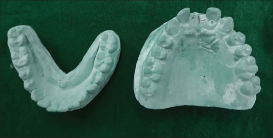
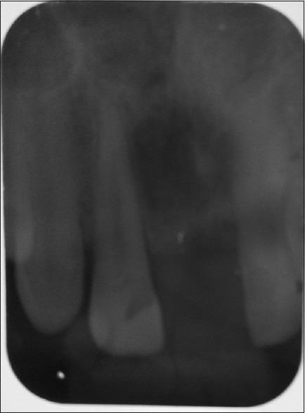
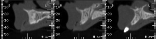
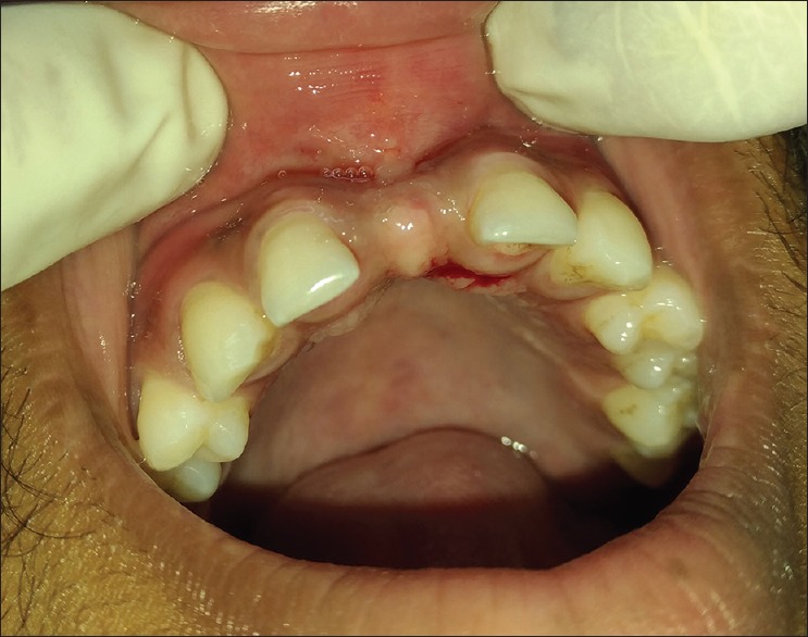
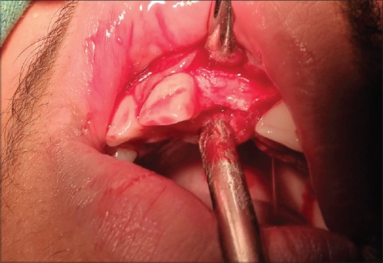
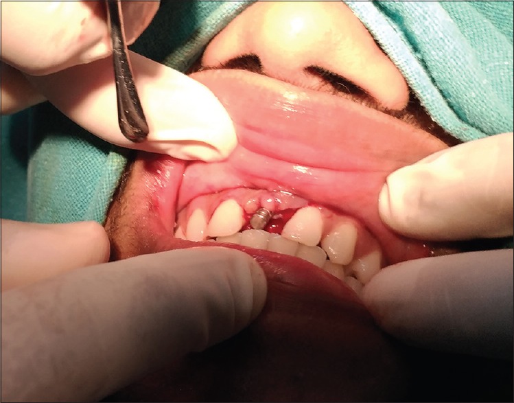
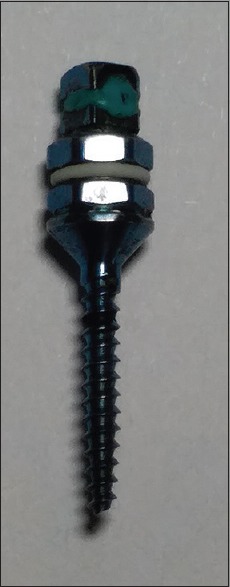
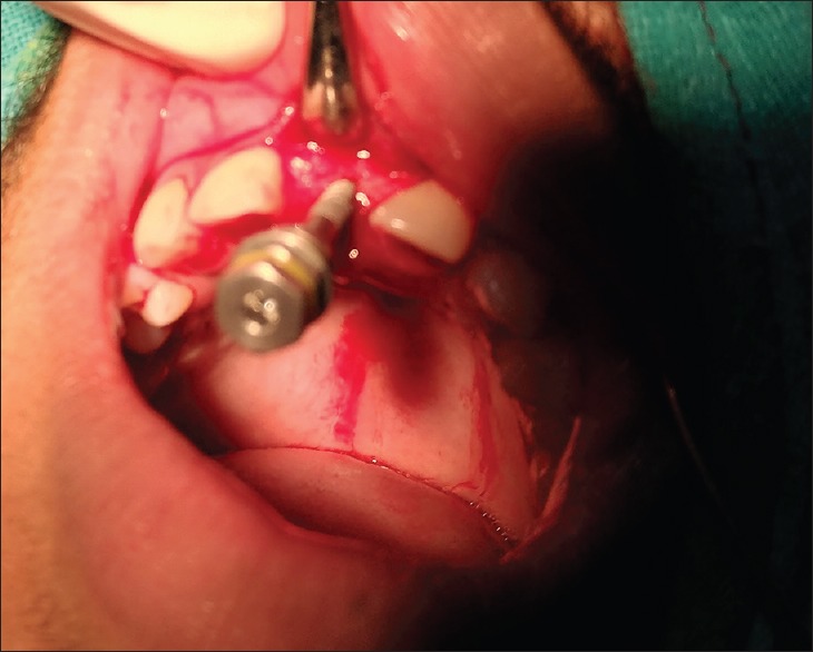
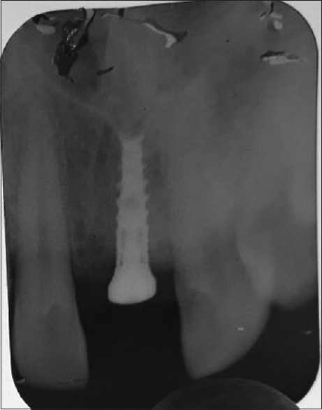
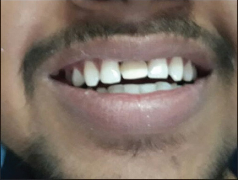
Implant placement is a challenge in the anterior maxilla if the available bone is reduced and esthetics is challenging. The ideal implant position should be considered in all three dimensions: mesiodistal, apicocoronal, and orofacial. This article includes a review and a case report for the anatomical and clinical perspective of implant placement in nasopalatine foramen (near incisal canal). In this case report, the edentulous space is mutilated in between the area #12 and #21 teeth. Therefore, only one, 3.0 W/10.00 L implant (bone size 4.2 mm width and 11 mm length) could be placed. Radiographically, D2 bone quality was diagnosed. Before surgery, an emphasis was given over the proper implant selection to avoid oversized implants due to critical anatomical landmark. Careful and with minimal trauma, the soft tissue was handled and implant placement was performed in a proper position, using information from panoramic radiograph, 3-D Dentascan. A surgical guide was used for placement of the implant. Finally, immediate loading of temporary implant prosthesis was done. The primary outcome was satisfactory, as after 72 h, no swelling and numbness were reported. The patient has been recalled after healing period of 24 weeks for permanent restoration.
Keywords: Bone expansion; dental implants; esthetics; imaging modalities; nasopalatine foramen region.
Conflict of interest statement
There are no conflicts of interest.
Figures


References
-
- Hoffmann KD. Anatomic considerations in the partially and fully edentulous maxilla. Atlas Oral Maxillofac Surg Clin North Am. 1994;2:31–9. - PubMed
-
- Buser D, Martin W, Belser UC. Optimizing esthetics for implant restorations in the anterior maxilla: Anatomic and surgical considerations. Int J Oral Maxillofac Implants. 2004;19(Suppl):43–61. - PubMed
-
- Bosse LP, Taylor TD. Problems associated with implant rehabilitation of the edentulous maxilla. Dent Clin North Am. 1998;42:117–27. - PubMed
-
- Carlsson GE, Bergman B, Hedegård B. Changes in contour of the maxillary alveolar process under immediate dentures. A longitudinal clinical and x-ray cephalometric study covering 5 years. Acta Odontol Scand. 1967;25:45–75. - PubMed
-
- Atwood DA. Some clinical factors related to rate of resorption of residual ridges. J Prosthet Dent. 1962;12:441–50. - PubMed
Publication types
LinkOut - more resources
Full Text Sources
Medical