
Strategic research agenda for biomedical imaging

European Institute for Biomedical Imaging Research (EIBIR). Insights Imaging. .

Abstract

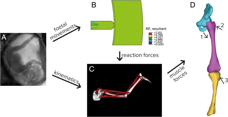
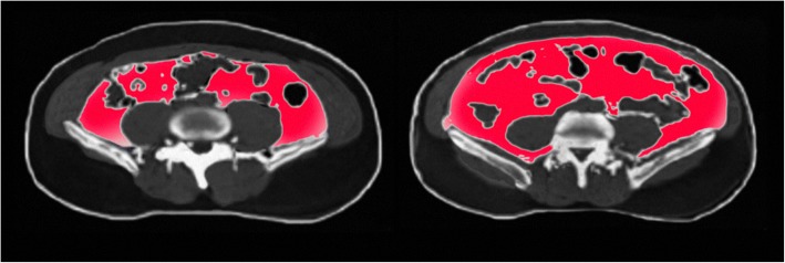
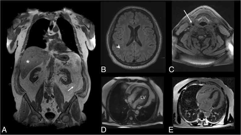
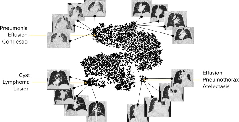
This Strategic Research Agenda identifies current challenges and needs in healthcare, illustrates how biomedical imaging and derived data can help to address these, and aims to stimulate dedicated research funding efforts.Medicine is currently moving towards a more tailored, patient-centric approach by providing personalised solutions for the individual patient. Innovation in biomedical imaging plays a key role in this process as it addresses the current needs for individualised prevention, treatment, therapy response monitoring, and image-guided surgery.The use of non-invasive biomarkers facilitates better therapy prediction and monitoring, leading to improved patient outcomes. Innovative diagnostic imaging technologies provide information about disease characteristics which, coupled with biological, genetic and -omics data, will contribute to an individualised diagnosis and therapy approach.In the emerging field of theranostics, imaging tools together with therapeutic agents enable the selection of best treatments and allow tailored therapeutic interventions.For prenatal monitoring, the use of innovative imaging technologies can ensure an early detection of malfunctions or disease.The application of biomedical imaging for diagnosis and management of lifestyle-induced diseases will help to avoid disease development through lifestyle changes.Artificial intelligence and machine learning in imaging will facilitate the improvement of image interpretation and lead to better disease prediction and therapy planning.As biomedical imaging technologies and analysis of existing imaging data provide solutions to current challenges and needs in healthcare, appropriate funding for dedicated research is needed to implement the innovative approaches for the wellbeing of citizens and patients.

Keywords: Artificial intelligence; Diagnostic imaging; Precision medicine; Preventive medicine; Radiology.


Conflict of interest statement

Competing interests

The author declares no competing interests.

Publisher’s Note

Springer Nature remains neutral with regard to jurisdictional claims in published maps and institutional affiliations.

Figures

Fig. 1
Fig. 1
MRI scan of early-stage 1A high-grade serous ovarian cancer. The BRCA1-positive patient was identified through ultrasound screening, confirmed by MRI. The right ovary contains as cyst with an enhancing nodule (red arrows) demonstrated on the T1 fat-saturated image before (a) and after (b) contrast administration. The nodule enhances brightly, similar to the adjacent uterine myometrium (white arrow). Detection of ovarian cancer at this early stage is associated with a significantly improved 5-year survival rate compared with later stages of disease (courtesy: A. Rockall)
Fig. 2
Fig. 2
ac Example of a 24-year-old female patient, with oligodendroglioma (grade II) in the left frontal lobe. a Cerebral blood flow, imaged by ASL. Red indicates high blood flow. b T1-Gd and c FLAIR. df Example of a 63-year-old female patient with GBM (grade IV) in the right temporal lobe. d Cerebral blood flow, imaged by ASL. Red indicates high blood flow. e T1 post Gd. f FLAIR. A clear difference in tumour blood flow can be seen between both patients (courtesy: A. Alsaedi, S. Bisdas)
Fig. 3
Fig. 3
Theranostics in a patient with a metastatic pancreatic neuroendocrine tumour. a 68Ga-Dotatate-PET scan for staging. b SPECT evaluation after treatment. c End-of-treatment 68Ga-Dotatate-PET scan (courtesy of J. Kunikowska, Warsaw)
Fig. 4
Fig. 4
Fluorescence imaging of a mouse showing fibroblast (red) migration from injection site to location of tumour cells (green). (a) Fibroblast recruitment to tumour injection site next to intensine. (b) Fibroblast recruitment to tumour patches attached to the peritoneal wall. (c) Fibroblast scattered in the abdomen of a control mouse without tumour injection. (d) High resolution image of fibroblasts recruited to a tumour. (e) experimental design (green) [12]
Fig. 5
Fig. 5
Computational pipeline for developing stress modelling of foetal movements. Foetal joint movements are tracked in utero (a), with finite element modelling of reaction forces (b) combined with musculoskeletal modelling to predict muscle forces (c) which are then applied to finite element models of foetal geometries (d). Adapted from [21]
Fig. 6
Fig. 6
BOLD imaging analysis of pregnant mice. Representative spatial distribution maps of the oxygen-haemoglobin dissociation inside the placenta and foetal liver on days 14.5 (a) and 17.5 (b) show distribution and variability [22]
Fig. 7
Fig. 7
Men matched for the same BMI and total body fat: Differing ‘adiposity phenotypes’ regarding visceral obesity and subcutaneous obesity and with different risk profiles (courtesy: A. Persson)
Fig. 8
Fig. 8
Imaging findings in a 61-year-old male indicating extensive subclinical disease burden. a Two-point DIXON T1-weighted sequence for the assessment of visceral adipose tissue volume from the femoral head to the cardiac apex (arrow) indicating high levels of fat as well as hepatic proton density fat fraction (asterisk, measured on multi-echo VIBE T1-weighted sequences). b Fluid-attenuated inversion recovery sequences demonstrating mild white matter lesions (arrowhead). c Atherosclerotic carotid plaque was determined on black-blood T1-weighted fat-suppressed sequences in the common carotid artery (arrow), the carotid bulb and the proximal internal carotid artery. d Cine-SSFP sequences were evaluated for the calculation of volume and mass left ventricle (LV). e late gadolinium enhancement was detected on fast-low-single-shot inversion recovery sequence four-chamber view [23]
Fig. 9
Fig. 9
The landscape of lung disease patients based on their CT image data. The distribution illustrates clusters of patients with similar imaging characteristics confirmed in reported findings. It is a step towards the identification of phenotypes in large-scale medical imaging data. [24]
Fig. 10
Fig. 10
Radiomics pipeline, with the aim to link imaging features to clinically relevant parameters such as tumour subtype, patient prognosis and therapy outcome prediction (Courtesy Martijn Starmans and Stefan Klein, Rotterdam)

References

    1. European Commission (2017) Shaping our Future. European Commission, Brussels Available via https://ec.europa.eu/info/events/shaping-our-future-2017-jul-03_en. Accessed 30 Aug 2018
    1. European Commission (2017) 2018 Commission work programme – key documents. European Commission, Brussels Available via https://ec.europa.eu/info/publications/2018-commission-work-programme-ke.... Accessed 30 Aug 2018
    1. European Commission . EU Council adopts conclusions on digital health care. Brussels: European Commission; 2017.
    1. Council of the European Union . Employment, Social Policy, Health and Consumer Affairs Council Meeting on 8 December 2017. 2017.
    1. European Society of Radiology (2011) Medical imaging in personalised medicine: a white paper of the research committee of the European Society of Radiology (ESR). Insights Imaging. 10.1007/s13244-011-0125-0 - PMC - PubMed

LinkOut - more resources