Review
. 2019 Mar;13(1):33-46.
doi: 10.1007/s12105-018-0977-4. Epub 2019 Jan 31.

A Guide to Yellow Oral Mucosal Entities: Etiology and Pathology

Affiliations
Review

A Guide to Yellow Oral Mucosal Entities: Etiology and Pathology

Duane R Schafer et al. Head Neck Pathol. 2019 Mar.

Abstract

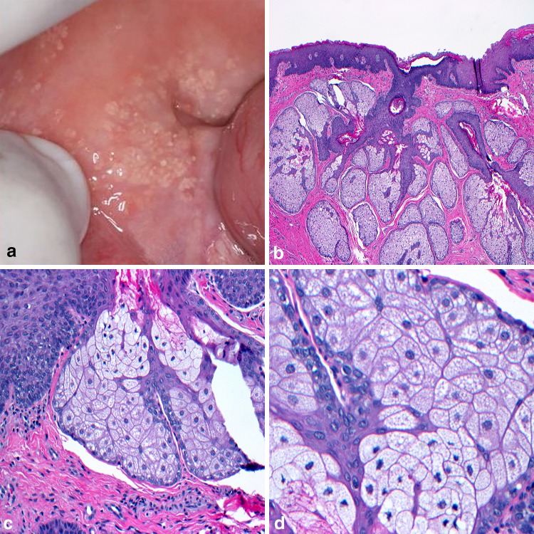
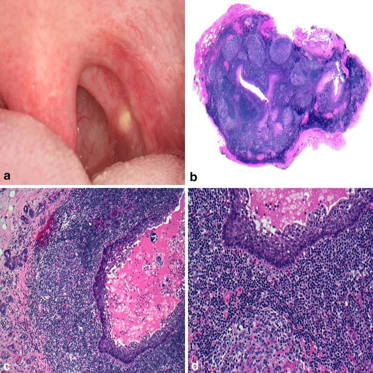
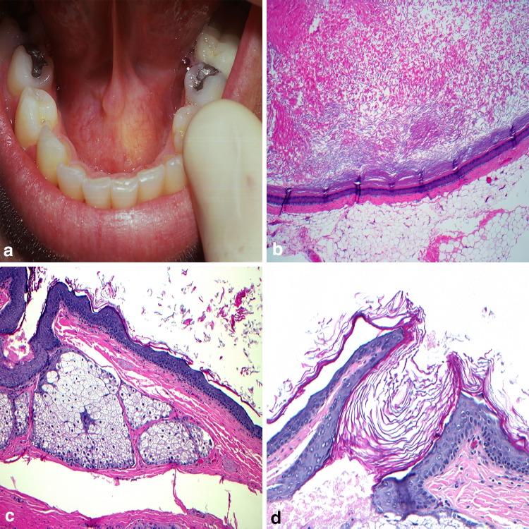
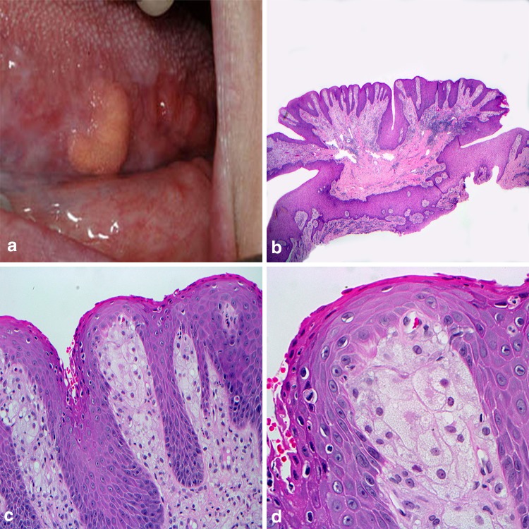
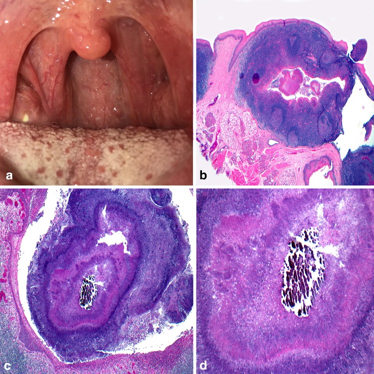
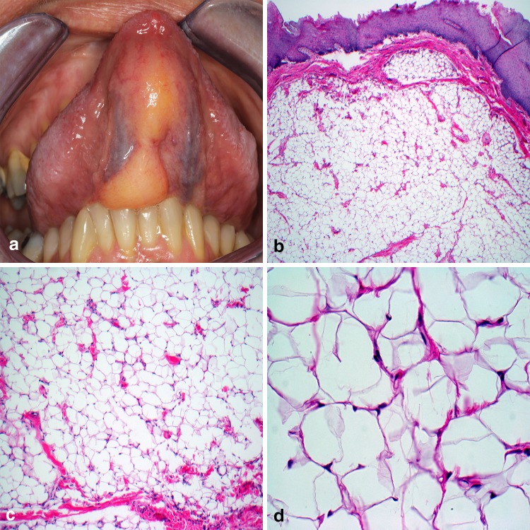
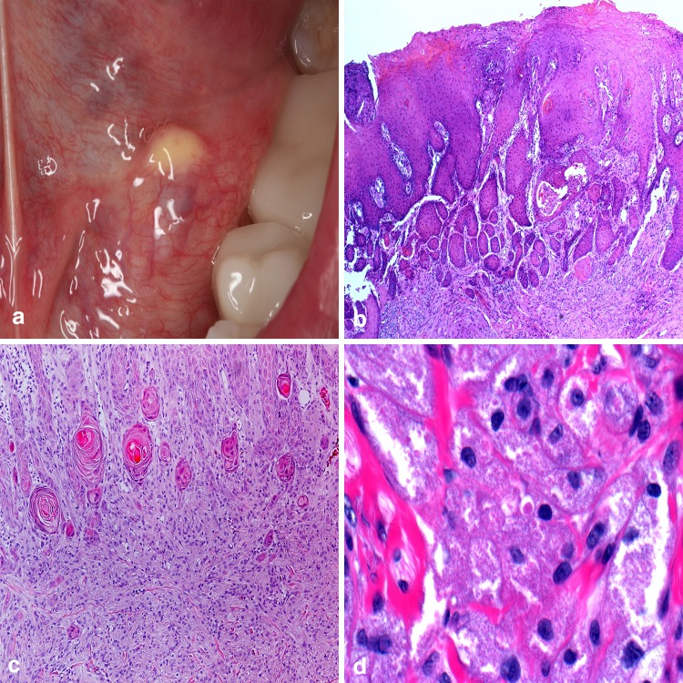
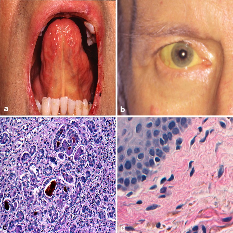
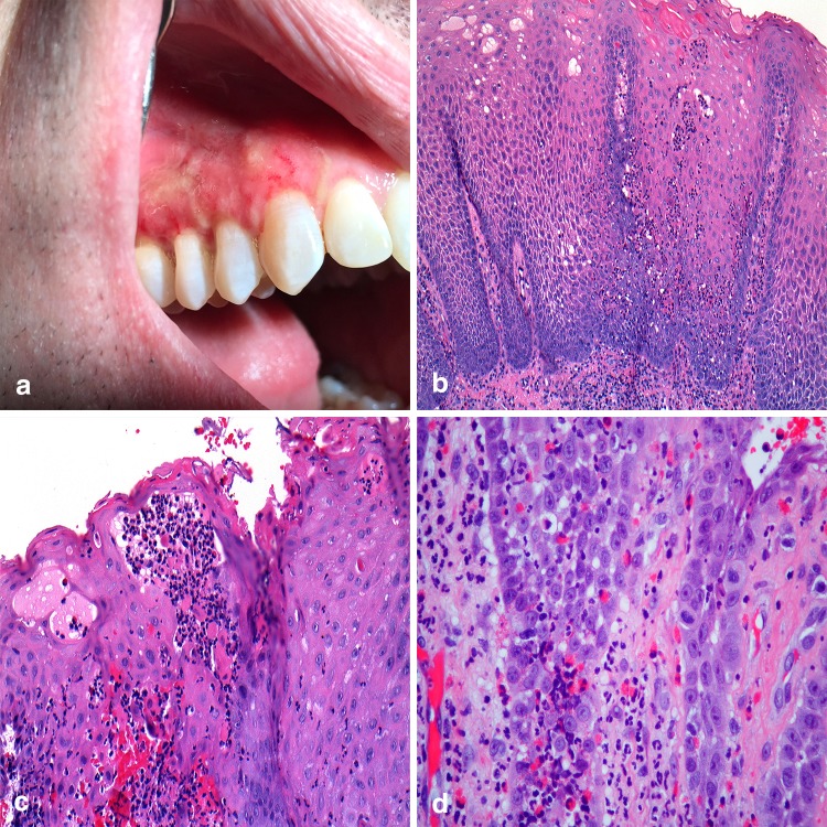
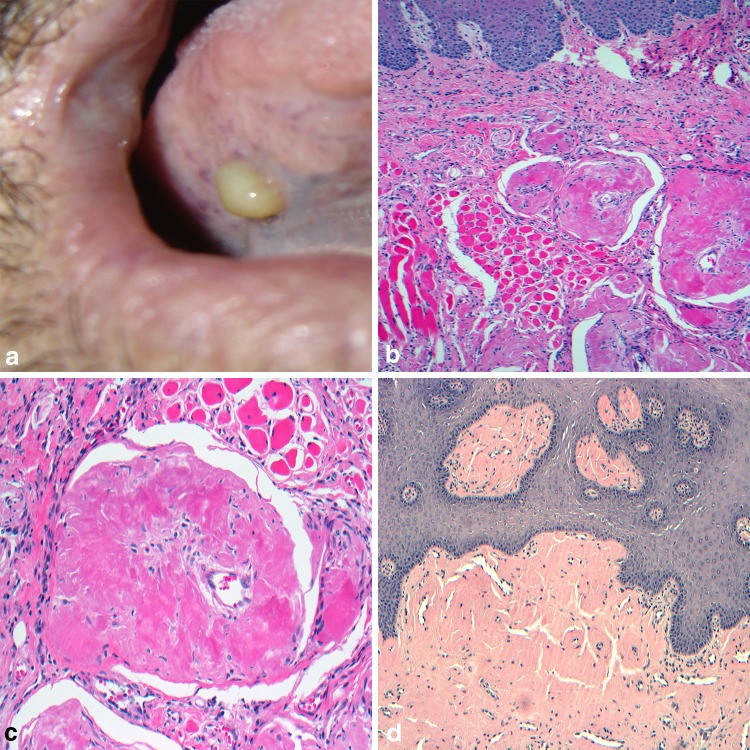
When faced with an uncertain clinical pathosis in the oral cavity, identifying the color of the mucosal lesion helps to narrow down a differential diagnosis. Although less common than red and white lesions, yellow lesions encompass a small group of distinct mucosal pathologic entities. Adipose tissue, lymphoid tissue, and sebaceous glands are naturally occurring yellow constituents of the oral cavity and become apparent with associated developmental or neoplastic lesions. Reactive and inflammatory lesions can create a yellow hue due to purulence, necrosis, and calcification. Some systemic diseases are known to deposit yellow bi-products such as amyloid or bilirubin into the oral mucosa of an affected person, and while not always yellow, unusual entities like verruciform xanthoma and granular cell tumor fall under the umbrella of yellow lesions given their occasional propensity to demonstration the color. This chapter aims to explore the unique group that is yellow lesions presenting in the oral mucosa.

Keywords: Mouth; Mucosal; Oral cavity; Oral manifestations; Systemic disease; Yellow.


Conflict of interest statement

Conflict of interest

All authors declare that they have no conflict of interest.

Ethical Approval

This article does not contain any studies with human participants or animals performed by any of the authors.

Informed Consent

As no human participants were involved in a study, informed consent was not required.

Figures

Fig. 1
Fig. 1
Fordyce granules. a Ectopic sebaceous glands distributed along the buccal mucosa; b lobular aggregate of sebaceous acinar units immediately below the mucosal epithelium; c intimate association of lobules with ductal elements; d large ovoid sebaceous cells with lipid filled vacuoles and a centrally placed nucleus
Fig. 2
Fig. 2
Oral lymphoepithelial cyst. a Slightly raised, yellow nodule arising in the tonsillar pillars; b squamous epithelial cyst lining surrounded by hyperplastic lymphoid tissue containing follicular pattern containing germinal centers; c desquamated epithelium and cellular debris filling the lumen; d lymphoid tissue abuts the cystic lining. Clinical photo courtesy of Dr. Kristin McNamara
Fig. 3
Fig. 3
Dermoid cyst. a Fluctuant swelling at midline of the floor of mouth; b orthokeratinized stratified squamous epithelial cyst lining with prominent keratohyalin granular cell layer; c sebaceous glands in the cyst wall; d dermal appendage elements intimately associated with the epithelium
Fig. 4
Fig. 4
Verruciform xanthoma. a Yellow-orange mass along the ventral lateral border of the tongue; b pebbly or warty surface architecture arising from surface mucosa; c parakeratinized hyperplastic epithelium with uniformly elongated rete pegs; d large polygonal, foamy-appearing, “xanthoma” cells fill the connective tissue papillae
Fig. 5
Fig. 5
Sialolithiasis. a Calcified “stone” within the submandibular gland excretory duct; b sialolith and inflammatory debris within the excretory duct lumen; c calcified mass appearing as concentric deposition; d occlusal radiograph confirms the presence of calcified mass. Clinical photo courtesy of Dr. Jerry Bouquot
Fig. 6
Fig. 6
Tonsillolith. a White-yellow mass noted protruding from a tonsillar crypt; b tonsillar crypt lined by stratified squamous epithelium and surrounded by reactive follicular hyperplasia; c crypt harboring concentrically organized bacterial colonies; d central calcified nidus surrounded by bacteria and neutrophils
Fig. 7
Fig. 7
Lipoma. a Soft, bosselated mass emanating from the ventral tongue; b well-circumscribed or encapsulated proliferation of mature adipose tissue; c thin fibrous strands containing inconspicuously scattered capillaries; d large vacuolated cells with peripherally displace nuclei. Clinical photo courtesy of Dr. Sarah Aguirre
Fig. 8
Fig. 8
Granular cell tumor. a Firm, yellow dome-shaped mass in the floor of the mouth; b overlying epithelium is nonulcerated and demonstrates pseudoepitheliomatous hyperplasia; c unencapsulated tumor cells arranged in ribbons and sheets around fibrous connective tissue septa and muscle; d large polygonal cells containing numerous autophagolysosomes imparting a course, gritty appearance to the cytoplasm. Clinical photo courtesy of Dr. Christine Harrington
Fig. 9
Fig. 9
Jaundice. a Diffuse yellow discoloration to the mucosa along the lingual frenum; b involvement of the sclera a common finding; c intraluminal plugging of ducts with bile with spillage into surrounding tissue; d peripheral tissues showing deposits along elastin fibers. Clinical photos courtesy Dr. Christopher Fielding
Fig. 10
Fig. 10
Pyostomatitis vegetans. a Irregular linear “snail” tracts along the maxillary alveolar mucosa; b papillary, acanthotic epithelium with inflammation; c intraepithelial microabscesses; d pronounced eosinophilia in lamina propria. Clinical photo courtesy of Dr. Paul Freedman
Fig. 11
Fig. 11
Amyloidosis. a Firm nodule on right ventral lateral tongue; b extracellular amorphous deposits of eosinophilic material in connective tissues; c affinity for muscle and small vessels; d highlighted by Congo red stain

Similar articles

Cited by

References

    1. Fordyce JA. A peculiar affection of the mucous membranes of the lips and the oral cavity. J Cutan Genito-Urin Dis. 1896;14:413–419.
    1. Neville BW, Damm DD, Allen CM, Chi AC. Developmental defects of the oral and maxillofacial region. Oral and maxillofacial pathology. 4. St. Louis: Elsevier; 2016. pp. 1–48.
    1. Batsakis JG, el-Naggar AK. Sebaceous lesion of salivary glands and oral cavity. Ann Otol Rhinol Laryngol. 1990;99:414–418. - PubMed
    1. Gaballah KY, Rahimi I, et al. Can presence of oral Fordyce’s granules serve as a marker for hyperlipidemia. Dent Res J. 2014;11:553–558. - PMC - PubMed
    1. Antonio N. Oral mucosa. Ten Cate’s oral histology. 9. St. Louis: Elsevier; 2017. pp. 260–288.