Review
. 2019 Mar;13(1):114-124.
doi: 10.1007/s12105-019-01005-5. Epub 2019 Jan 29.

Tongue Lumps and Bumps: Histopathological Dilemmas and Clues for Diagnosis

Affiliations
Review

Tongue Lumps and Bumps: Histopathological Dilemmas and Clues for Diagnosis

Irit Allon et al. Head Neck Pathol. 2019 Mar.

Abstract

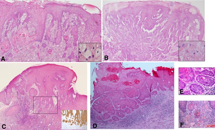
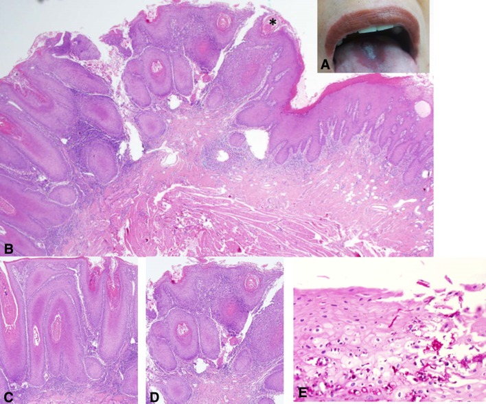
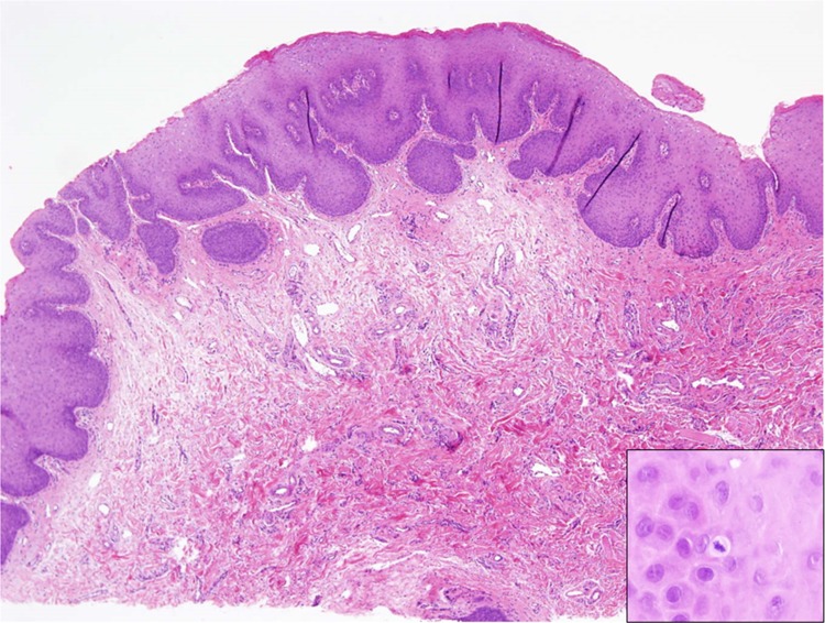
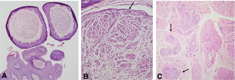
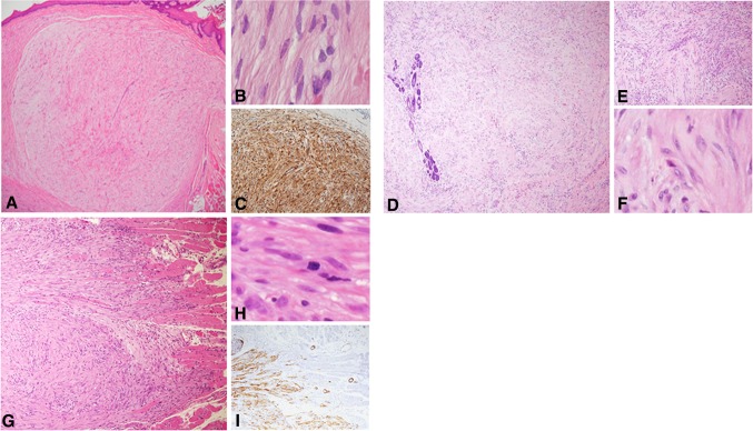
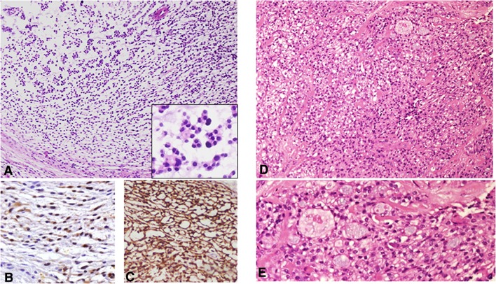
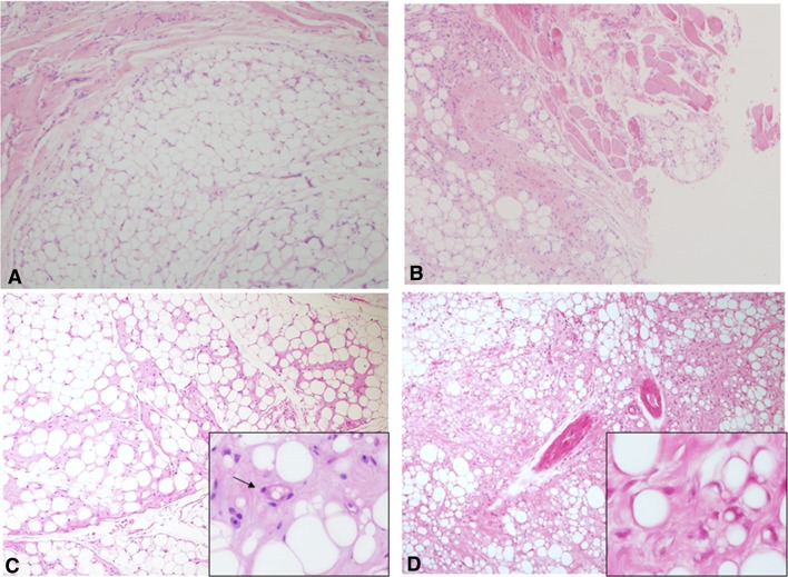
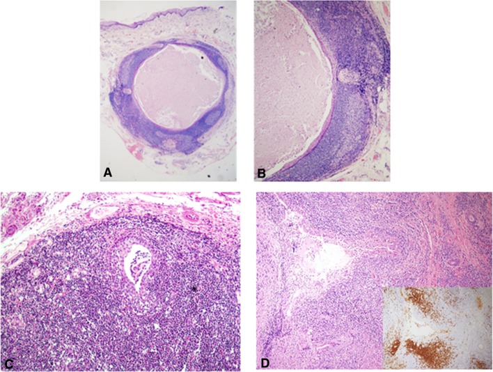
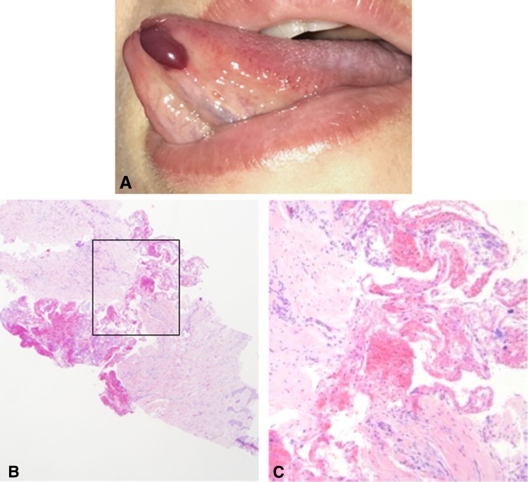
Exophytic lesions of the tongue encompass a diverse spectrum of entities. These are most commonly reactive, arising in response to local trauma but can also be neoplastic of epithelial, mesenchymal or miscellaneous origin. In most cases, the microscopic examination is likely to provide a straightforward diagnosis. However, some cases can still raise microscopic diagnostic dilemmas, such as conditions that mimic malignancies, benign tumors with overlapping features and anecdotal lesions. A series of "lumps and bumps" of the tongue are presented together with suggested clues that can assist in reaching a correct diagnosis, emphasizing the importance of the clinico-pathological correlations.

Keywords: Adipocytic tumors; Lumps and bumps; Lymphoid-rich lesions; Neural lesion; Pseudoepitheliomatous hyperplasia; Spindle cell tumors; Tongue; Verrucous lesions.


Conflict of interest statement

Conflict of interest

Irit Allon, Marilena Vered declares, Ilana Kaplan declares that they have no conflict of interest.

Research Involving Human Participants and/or Animals

This article does not contain any studies with human participants performed by any of the authors.

Figures

Fig. 1
Fig. 1
a Pseudoepitheliomatous hyperplasia of the lining oral epithelium associated with granular cell tumor. Deeply set and separate epithelial islands with keratin pearls are such a prominent feature that the adjacent granular cells may be overlooked (inset). This may lead to misdiagnosis of the case as well-differentiated squamous cell carcinoma. However, the hyperplasia lacks cellular atypia and represents a completely benign process (H&E, original magnification × 100). b An additional example of florid pseudoepitheliomatous hyperplasia associated with granular cell tumor mimicking infiltration, which may lead to misdiagnosis as squamous cell carcinoma. Granular cells are present but scarce (inset) (H&E, original magnification × 100). c In biopsy from the lateral aspect of the tongue, the distinction between pseudoepitheliomatous hyperplasia and well-differentiated squamous cell carcinoma necessitated immunostaining with cytokeratin that highlighted frank infiltration in the form of small epithelial clusters and nests (inset), leading to the diagnosis of squamous cell carcinoma. (H&E, original magnification × 40). d Another disputable lesion of the lateral tongue on the background of oral lichen planus. (H&E, original magnification × 100). e and f The malorientation of the specimen preventing accurate diagnosis was solved with serial deep sections in which true invasion was demonstrated (H&E, e original magnification × 200, f original magnification × 400)
Fig. 2
Fig. 2
a A white exophytic, nodular symmetrical lesion at the midline dorsal aspect of the tongue, middle third. An erythematous, smooth-surfaced hallow surrounds the white lesion. Although a diagnosis of median rhomboid glossitis has been clinically submitted, an incisional biopsy was taken due to the unusual appearance of this particular lesion. b Microscopically, the lesion demonstrated a nodular-to-verrucous type of epithelial proliferation, hyperkeratosis, including formation of keratin “plugs” and an associated band of chronic inflammation, all of which confer the lesion an appearance reminiscent of verrucous carcinoma (H&E, original magnification × 40). c and d A higher magnification shows no remarkable cellular atypia (H&E, original magnification × 100) a common feature to both MRG and VC. e Candidal hyphae are seen penetrating the surface (PAS, original magnification × 600). The clinical and microscopical findings favored a diagnosis of “nodular” median rhomboid glossitis over that of verrucous carcinoma
Fig. 3
Fig. 3
A biopsy from a girl with multiple exophytic lesions on the oral mucosa including the tongue. Each lesion is about 0.5 cm in diameter. Microscopically, there is hyperplasia and acanthosis of the lining epithelium overlying an exuberant collagenous connective tissue (H&E, original magnification × 40). The differential diagnosis was between multiple lesions of reactive fibrous hyperplasia (i.e., fibrous epulis/irritation fibroma) and Heck’s disease. The identification of a mitosoid cell in the spinous layer (inset) favored a diagnosis of Heck’s disease
Fig. 4
Fig. 4
a This is a biopsy from a 30 years old male with multiple pale raised papules on the anterior dorsal surface of the tongue with a yet undiagnosed MEN2B. Microscopically, there are subepithelial, well-circumscribed spindle cell nodules (H&E, original magnification × 40). b At a higher magnification, the nodules contain axonal-like structures embedded in a background of loose connective tissue, with a delicate capsule at the periphery (arrow) (H&E, original magnification × 200). The diagnosis was multiple mucosal neuromas, which demands a general work-up to rule out the diagnosis of MEN2B syndrome. The capsule of the nodules represents the perineurium. This patient was diagnosed with a medullary carcinoma of the thyroid. c This is an example of a traumatic neuroma from the tongue in a 31 years old male patient. Clinically, the lesion was mildly painful and appeared following a previous biopsy in the same area. Note the unusual thickened perineurium around the nerve bundles (arrows) (H&E, original magnification × 100)
Fig. 5
Fig. 5
a Leiomyoma of the tongue shows a well-circumscribed highly cellular lesion with intersecting bundles of spindle-shaped cells (H&E, original magnification × 40). b “Cigar”-shaped nuclei of the cells suggested leiomyoma (H&E, original magnification × 400). c Immunostaining with h-caldesmon shows a diffuse and strong immunoreaction (h- caldesmon, original magnification × 100). d Myofibroma of the tongue presenting a highly cellular lesion with intersecting bundles of spindle-shaped cells involving the salivary glands of the posterior tongue (H&E, original magnification × 40). e The-zonation phenomenon of interchangeable darker and lighter areas of myofibroblastic cells is typical for myofibroma (H&E, original magnification × 100). f The nuclei of myofibroblasts have crescent sharp edges (H&E, original magnification × 200). g Low-grade myofibroblastic sarcoma infiltrating into the tongue striated muscles (H&E, original magnification × 40). h Cellular atypia in low-grade myofibroblastic sarcoma; a mitotic figure in the center of the photomicrograph (H&E, original magnification × 400). i Calponin immunoreactivity in low-grade myofibroblastic sarcoma highlights the infiltrative pattern of the tumor (Calponin, original magnification × 40)
Fig. 6
Fig. 6
a Microscopically, ectomesenchymal chondro-myxoid tumor is usually characterized by a lobulated architecture with a myxoid stroma and bland polygonal cells arranged in a globoid pattern (H&E, original magnification × 40). Inset: tumor cells with eosinophilic cytoplasm and dark staining nuclei. b S100 is typically positive in ectomesenchymal chondro-myxoid tumor (S100, original magnification × 100). c GFAP positivity in ectomesenchymal chondro-myxoid tumor (GFAP, original magnification × 100). d Clear cell carcinoma of salivary glands can also present nests of monomorphic clear cells in a myxoid-to-hyaline stroma and a glandular-like arrangement, which occasionally could raise the differential diagnosis with ectomesenchymal chondro-myxoid tumor (H&E, original magnification × 100). e Clear cells, myxoid areas and a glandular-like pattern of clear cell carcinoma (H&E, original magnification × 200)
Fig. 7
Fig. 7
a Lipoma of the tongue showing a well circumscribed tumor of mature, uniformly sized adipocytes (H&E, original magnification × 40). b Intramuscular lipoma features an infiltrative growth pattern of mature adipocytes between the muscle fibers (H&E, original magnification × 40). c Lipoma with lipoblasts is essentially a lipoma same as in a, however occasional cells resembling lipoblasts can be encountered (inset) (H&E, original magnification × 40). d Atypical lipomatpus tumor/well-differentiated liposarcoma of the tongue presents an infiltrative growth pattern, variably sized adipocytes and fibrous septae containing atypical neoplastic adipocytes (H&E, original magnification × 40). Inset shows some multivacuolar lipoblasts with eccentric, crescent/signet-ring-like nuclei
Fig. 8
Fig. 8
a A small, intramucosal cystic lesion from the ventral surface of the tongue (H&E, original magnification × 40). b The rim of lymphoid tissue, including germinal centers, is a prominent feature (H&E, original magnification × 200). This was diagnosed as a lymphoepithelial cyst. c A dilated and hyperplastic minor salivary gland duct is surrounded, and occasionally encroached, by a massive lymphocytic infiltrate. Some of the inflammatory cells have the appearance of monocytoid lymphocytes with a clear halo. These features are reminiscent of lympho-epithelial lesions (H&E, original magnification × 40). d A similar finding as in c, however, in this case the monocytoid lymphocytes invaded extensively into the salivary gland epithelium. These lymphocytes were CD20 positive (inset). Polymerase chain reaction demonstrated an IgD kappa monoclonal lymphocytic population supporting the diagnosis of extranodal marginal zone B cell lymphoma (H&E, original magnification × 40)
Fig. 9
Fig. 9
a A blood-filled bulla at the anterior ventral tongue can be clinically diagnosed as angina bullosa hemorrhagica, given that there are no coagulation deficiencies or anti-coagulant medications. b Microscopically, there is lack of epithelial lining, which might lead to an erroneous diagnosis of a vesiculo-bullous disease characterized by sub-epithelial split with loss of the oral epithelium (H&E, original magnification × 40). c At a higher magnification, hemorrhagic areas can be observed (H&E, original magnification × 200)

References

    1. Brad W, Neville DD, Chi AC, Allen CM. Oral and maxillofacial pathology, 4th edn. Elsevier Health Sciences; 2015. p. 34–35, 336–338, 389–391, 473–475, 497–498, 502–503, 512–514.
    1. Awadallah M, Idle M, Patel K, Kademani D. Management update of potentially premalignant oral epithelial lesions. Oral Surg Oral Med Oral Pathol Oral Radiol. 2018;125:628–636. doi: 10.1016/j.oooo.2018.03.010. - DOI - PubMed
    1. Angiero F, Berenzi A, Benetti A, Rossi E, Del Sordo R, Sidoni A, Stefani M, Dessy E. Expression of p16, p53 and Ki-67 proteins in the progression of epithelial dysplasia of the oral cavity. Anticancer Res. 2008;28:2535–2539. - PubMed
    1. Zarovnaya E, Black C. Distinguishing pseudoepitheliomatous hyperplasia from squamous cell carcinoma in mucosal biopsy specimens from the head and neck. Arch Pathol Lab Med. 2005;129:e1032-6. - PubMed
    1. Hosseinpour S, Mashhadiabbas F, Ahsaie MG. Diagnostic biomarkers in oral verrucous carcinoma: a systematic review. Pathol Oncol Res. 2017;23:19–32. doi: 10.1007/s12253-016-0150-x. - DOI - PubMed