Case Reports
. 2019 Mar;7(3):e566.
doi: 10.1002/mgg3.566. Epub 2019 Jan 28.

Protein informatics combined with multiple data sources enriches the clinical characterization of novel TRPV4 variant causing an intermediate skeletal dysplasia

Affiliations
Case Reports

Protein informatics combined with multiple data sources enriches the clinical characterization of novel TRPV4 variant causing an intermediate skeletal dysplasia

Stephanie L Hines et al. Mol Genet Genomic Med. 2019 Mar.

Abstract

Background: Transient receptor potential cation channel subfamily V member 4 (TRPV4) is an ion channel permeable to Ca2+ that is sensitive to physical, hormonal, and chemical stimuli. This protein is expressed in many cell types, including osteoclasts, chondrocytes, and sensory neurons. As such, pathogenic variants of this gene are associated with skeletal dysplasias and neuromuscular disorders. Pathogenesis of these phenotypes is not yet completely understood, but it is known that genotype-phenotype correlations for TRPV4 pathogenic variants often are not present.

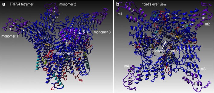
Methods: Newly characterized, suspected pathogenic variant in TRPV4 was analyzed using protein informatics and personalized protein-level molecular studies, genomic exome analysis, and clinical study.

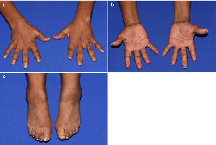
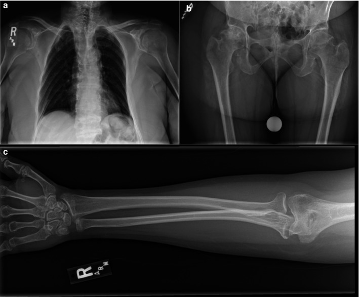
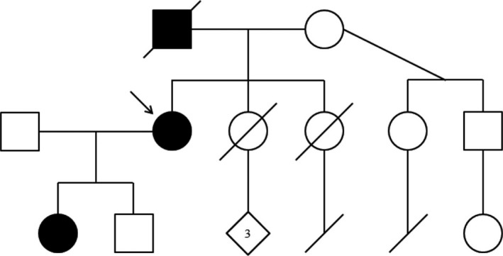
Results: This statement is demonstrated in the family of our proband, a 47-year-old female having the novel c.2401A>G (p.K801E) variant of TRPV4. We discuss the common symptoms between the proband, her father, and her daughter, and compare her phenotype to known TRPV4-associated skeletal dysplasias.

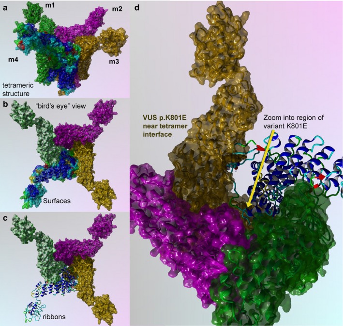
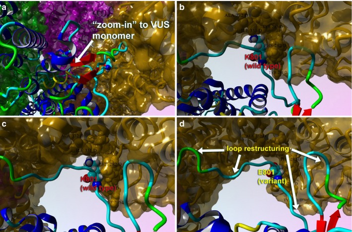
Conclusions: Protein informatics and molecular modeling are used to confirm the pathogenicity of the unique TRPV4 variant found in this family. Multiple data were combined in a comprehensive manner to give complete overall perspective on the patient disease and prognosis.

Keywords: Kozlowski type; Maroteaux type; skeletal dysplasia; spondyloepiphyseal dysplasia; spondylometaphyseal dysplasia; transient receptor potential cation channel subfamily V member 4 (TRPV4).


Conflict of interest statement

All authors declare that they have no conflict of interest.

Figures

Figure 1
Figure 1
Physical abnormalities in the proband. Multiple digit abnormalities are visualized, including widened PIPs, brachydactyly, and hypoplastic nails. (a) Front of hands. (b) Reverse side of hands. (c) Feet are notable for reduced size, brachydactyly, and hypoplastic nails
Figure 2
Figure 2
X‐ray images of the proband. (a) Thoracic abnormalities as seen through X‐ray, note platyspondyly, asymmetry of ribs, and absence of scoliosis. (b) X‐ray of the hips, note reduced joint space between hip and right femur. (c) Right arm/hand of proband, metaphyseal abnormality is visualized
Figure 3
Figure 3
Pedigree of the proband's family. Individuals possessing skeletal abnormalities have been colored black. Note the autosomal‐dominant inheritance pattern—individuals without skeletal abnormalities have children also lacking skeletal abnormality. An arrow marks the proband
Figure 4
Figure 4
TRPV4 wild‐type structure for full‐length human sequence consisting of 871 amino acids. (a) Full‐length model for the entire TRPV4 structure that shows four monomer proteins that comprise the cation channel, as depicted in ribbons. Colors indicate the composite technique for our hybrid model technology to generate highly reliable structural models for VUS determination. This structural model is complete full‐length sequence including residues and loops missing from X‐ray structures. (b) Rotated view to look down into the channel pore, called the “bird's eye” view. The labeling for both models is indicated. Residues shown are rendered in either “licorice sticks” or van Der Waals (VdW) and using standard element coloring (O‐red, N‐blue, H‐white, S‐yellow) except for the carbon atoms that are colored to match the ribbon color
Figure 5
Figure 5
TRPV4 surface and mapping for interaction for wild type and p.K801E variant. (a) Full‐length model for the entire TRPV4 tetrameric structure colored to distinguish the four protein monomer chains. Monomers 1–3 are colored uniformly, and monomer 4 is colored by secondary structure. (b) Bird's eye view for the wild‐type surface rendered model is shown. (c) Key region to zoom into is shown in ribbons and colored by secondary structure. (d) Zoom by 5X and rotation by the X‐Y plane at 135° are done to show position of the VUS (p.K801E), which has a critical point located near the center of the tetramer interface
Figure 6
Figure 6
Close‐up views of the TRPV4 variant (p.K801E) surface and variant effect on loop structure and effect on tetrameric interface. (a) Full‐length model for the variant TRPV4 tetrameric structure colored by secondary structure and atom type. Interacting residues between the two proteins are shown. (b) The tetrameric structure for wild type (K801) is shown with same scheme. (c) Close‐up view of the loops is shown for the wild type in this monomer–monomer interaction region. (d) Same view for the p.K801E is shown to illustrate the changes in the loop conformation and the effect on tetramer conformation. This VUS causes the pore structure to be altered for the cation channel, which also demonstrates changes in the protein Gibbs‐free energy for stability

References

    1. Abdul‐Hay, S. O. , Lane, A. L. , Caulfield, T. R. , Claussin, C. , Bertrand, J. , Masson, A. , … Leissring, M. A. (2013). Optimization of peptide hydroxamate inhibitors of insulin‐degrading enzyme reveals marked substrate‐selectivity. Journal of Medicinal Chemistry, 56(6), 2246–2255. 10.1021/jm301280p - DOI - PMC - PubMed
    1. Altschul, S. F. , Madden, T. L. , Schäffer, A. A. , Zhang, J. , Zhang, Z. , Miller, W. , & Lipman, D. J. (1997). Gapped BLAST and PSI‐BLAST: a new generation of protein database search programs. Nucleic Acids Research, 25(17), 3389–3402. 10.1093/nar/25.17.3389 - DOI - PMC - PubMed
    1. Ando, M. , Fiesel, F. C. , Hudec, R. , Caulfield, T. R. , Ogaki, K. , Górka‐Skoczylas, P. , … Springer, W. (2017). The PINK1 I368N mutation affects protein stability and ubiquitin kinase activity. Molecular Neurodegeneration, 12(1), 32 10.1186/s13024-017-0174-z - DOI - PMC - PubMed
    1. Caulfield, T. R. (2011). Inter‐ring rotation of apolipoprotein A‐I protein monomers for the double‐belt model using biased molecular dynamics. Journal of Molecular Graphics and Modelling, 29(8), 1006–1014. 10.1016/j.jmgm.2011.04.005 - DOI - PubMed
    1. Caulfield, T. , & Devkota, B. (2012). Motion of transfer RNA from the A/T state into the A‐site using docking and simulations. Proteins, 80(11), 2489–2500. 10.1002/prot.24131 - DOI - PubMed

Publication types