

Postmortem magnetic resonance imaging vs autopsy of second trimester fetuses terminated due to anomalies
- PMID: 30694559
- PMCID: PMC6618902
- DOI: 10.1111/aogs.13548
Postmortem magnetic resonance imaging vs autopsy of second trimester fetuses terminated due to anomalies
Abstract
Introduction: Our aim was to investigate the accuracy of postmortem fetal magnetic resonance imaging (MRI) compared with fetal autopsy in second trimester pregnancies terminated due to fetal anomalies. A secondary aim was to compare the MRI evaluations of two senior radiologists.
Material and methods: This was a prospective study including 34 fetuses from pregnancies terminated in the second trimester due to fetal anomalies. All women accepted a postmortem MRI and an autopsy of the fetus. Two senior radiologists performed independent evaluations of the MRI images. A senior pathologist performed the fetal autopsies. The degree of concordance between the MRI evaluations and the autopsy reports was estimated as well as the consensus between the radiologists.
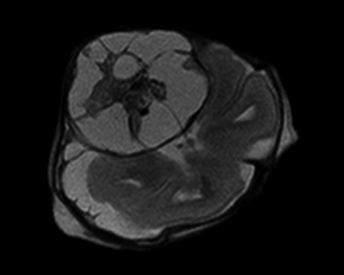
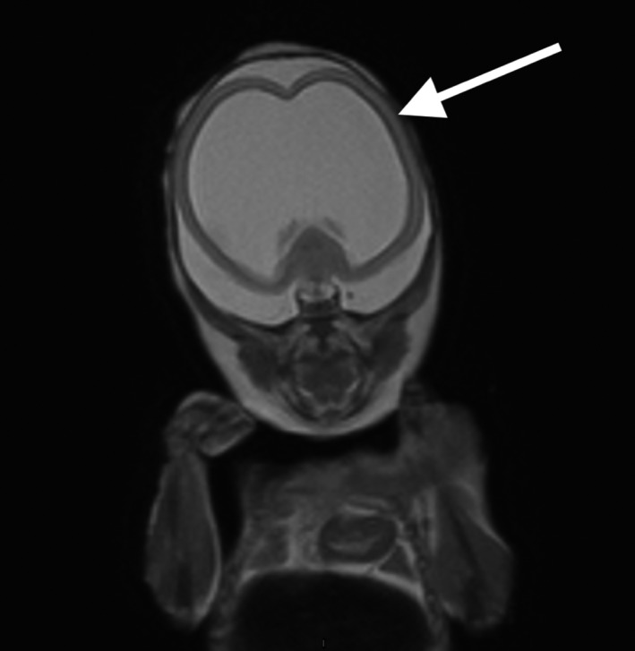
Results: Thirty-four fetuses were evaluated. Sixteen cases were associated with the central nervous system (CNS), five were musculoskeletal, one cardiovascular, one was associated with the urinary tract, and 11 cases had miscellaneous anomalies such as chromosomal aberrations, infections and syndromes. In the 16 cases related to the CNS, both radiologists reported all or some, including the most clinically significant anomalies in 15 (94%; CI 70%-100%) cases. In the 18 non-CNS cases, both radiologists reported all or some, including the most clinically significant anomalies in six (33%; CI 5%-85%) cases. In 21 cases (62%; CI 44%-78%), both radiologists held opinions that were consistent with the autopsy reports. The degree of agreement between the radiologists was high, with a Cohen's Kappa of 0.87.
Conclusions: Postmortem fetal MRI can replace autopsy for second trimester fetuses with CNS anomalies. For non-CNS anomalies, the concordance is lower but postmortem MRI can still be of value when autopsy is not an option.
Keywords: fetal anomalies; fetal diagnosis; postmortem fetal MRI; prenatal diagnosis; prospective study; second trimester.
© 2019 The Authors. Acta Obstetricia et Gynecologica Scandinavica published by John Wiley & Sons Ltd on behalf of Nordic Federation of Societies of Obstetrics and Gynecology (NFOG).
Conflict of interest statement
None of the authors report any conflicts of interest.
Figures


References
-
- SBU . The Swedish Council on Technology assessment in Health Care. Routine ultrasound under pregnancy, report. Stockholm: ISBU, 1998.
-
- Amini H, Antonsson P, Papadogiannakis N, et al. Comparison of ultrasound and autopsy findings in pregnancies terminated due to fetal anomalies. Acta Obstet Gynecol Scand. 2006;85(10):1208‐1216. - PubMed
-
- Dickinson JE, Prime DK, Charles AK. The role of autopsy following pregnancy termination for fetal abnormality. Aust N Z J Obstet Gynaecol. 2007;47(6):445‐449. - PubMed
Publication types
MeSH terms
Grants and funding
LinkOut - more resources
Full Text Sources
Medical
