Review
. 2019 Jun;130(6):1049-1063.
doi: 10.1097/ALN.0000000000002554.

Understanding the Effects of General Anesthetics on Cortical Network Activity Using Ex Vivo Preparations

Affiliations
Review

Understanding the Effects of General Anesthetics on Cortical Network Activity Using Ex Vivo Preparations

Logan J Voss et al. Anesthesiology. 2019 Jun.

Abstract

General anesthetics have been used to ablate consciousness during surgery for more than 150 yr. Despite significant advances in our understanding of their molecular-level pharmacologic effects, comparatively little is known about how anesthetics alter brain dynamics to cause unconsciousness. Consequently, while anesthesia practice is now routine and safe, there are many vagaries that remain unexplained. In this paper, the authors review the evidence that cortical network activity is particularly sensitive to general anesthetics, and suggest that disruption to communication in, and/or among, cortical brain regions is a common mechanism of anesthesia that ultimately produces loss of consciousness. The authors review data from acute brain slices and organotypic cultures showing that anesthetics with differing molecular mechanisms of action share in common the ability to impair neurophysiologic communication. While many questions remain, together, ex vivo and in vivo investigations suggest that a unified understanding of both clinical anesthesia and the neural basis of consciousness is attainable.


Figures

Figure 1.
Figure 1.. Ex vivo slice preparations of the brain and common recording configurations in neocortex.
(A) schematic illustrating preparation of brain slices. Brains are sliced in any desirable plane and orientation (shown is a coronal slicing plane) and, depending on subsequent use, the slices may be trimmed to include just the region of interest. The resulting slices are either allowed to recover 1-2 h and are then used for experimentation on the same day ('acute slice') or are placed on a substrate and cultivated in nutritional medium, resulting in an organotypic slice culture. Acute slices can be prepared from animals of any age (commonly juveniles) whereas for cultures neonatal animals or even embryos are often required. (B) sketch of a partial coronal brain slice including neocortex, thalamus and hippocampus. Blue dots illustrate commonly used orientations of multi-channel recording sites within neocortex. In the 'horizontal' orientation the sites are situated within one layer or along layer boundaries, allowing the recording of inter-areal propagation of neuronal activity. The 'vertical', cross-layer orientation, running parallel to pyramidal cells' apical dendrites, is usually chosen if the spread of activity within a cortical column/across layers is of interest, e.g. upon thalamic stimulation. Two pyramidal cells (magenta) are shown schematically to illustrate the orientation of the long apical dendrites.
Figure 2.
Figure 2.. Pathway specificity of isoflurane effects in auditory cortex.
(A, B): Current source density responses to cortico-cortical (A; CC; ‘top-down’) and thalamo-cortical (B; TC; ‘bottom-up’) synaptic responses in murine brain slices of primary auditory cortex in control (left column) and recovery (right column) and three doses of isoflurane (middle columns). In each panel, vertical axis corresponds to normalized cortical depth (pial surface at top, white matter at the bottom). Horizontal grey lines indicate cortical layer boundaries. Blue colors correspond to current sinks, i.e. excitatory synaptic currents flowing into cells. (C): Magnitude of layer ¾ TC sink (red) and layer 1 CC sink (blue) from the data in A, showing greater suppression by isoflurane of CC responses compared to TC. (D): Same as C but showing the 2-D cross-correlation between sink pattern at each drug condition with the pattern in control. Reproduced under the terms of the Creative Commons Attribution Licence from .
Figure 3.
Figure 3.. Summary of effects of general anesthetics on long range connectivity in the cortico-thalamic network.
Schematic showing major feedforward and feedback afferent pathways in the cortico-thalamic network. Under awake conditions (left), projections from ‘core’ cells (blue) in thalamus carry specific information to granular layers (L4) in neocortex, while ‘matrix’ cells (red) exert modulatory influences in supragranular and infragranular layers (L1, L5). Feedforward cortical projection cells (cyan) in supragranular layers (L2/3) project to higher order cortex, while feedback cortical projection cells (magenta) in infragranular layers (L5/6) project to lower order cortex (and subcortically; not shown). Under doses of anesthesia causing LOC (right), feedback cortico-cortical and matrix thalamo-cortical projections are suppressed relative to feedforward cortico-cortical and core thalamo-cortical projections.
Figure 4.
Figure 4.. Isoflurane depresses polysynaptic bursts more than monosynaptically driven activity.
(A) Current source density (CSD) plots of activity in a mouse auditory thalamocortical slice induced by electrical stimulation of thalamic afferents. The vertical extent of the plots spans the entire cortical depth. Arrowheads indicate the times of occurrence of the stimulation pulses (four pulses at 40 Hz). Cold colors represent current sinks, warm colors current sources. Brief monosynaptic responses (~10 ms) appear immediately after each stimulation pulse, whereas the much longer bursts arise after the third stimulation pulse and evolve over hundreds of milliseconds post-stimulus. Compare the almost complete depression of bursts by isoflurane to the moderate attenuation of the monosynaptic responses. (B) Depression of monosynaptically driven ('early') spiking activity in thalamocortical slices by isoflurane. Each point represents the integral of these early responses (see for details) from a slice, normalized to the drug-free condition. TC denotes thalamocortical stimulation. (C) Integral of burst activity induced by TC and cortical layer 1 (L1) stimuli (same conventions as in B apply). Reproduced under the terms of the Creative Commons Attribution License. Panel A modified from ; panels B-C slightly modified from .
Figure 5.
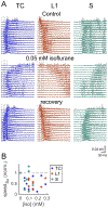
Figure 5.. Anesthetics slow and impair propagation of cortical activity in acute thalamo-cortical slices.
(A) Example of neocortical burst activity in a thalamocortical slice. Bursts were either induced by electrical stimuli (dotted lines) in auditory thalamus (TC) or in cortical layer 1 (L1) or arose spontaneously; they were extracellularly recorded from a linear 16 channel-array placed in layer 5 of neocortex. Gray traces are three representative trials; colored thick traces are averages. Note the speedy uni- or bidirectional burst propagation during control, and its impairment by a very small concentration of isoflurane. (B) Speed of burst propagation in various isoflurane concentrations, normalized to control. Each filled circle is one slice. Modified and reproduced under the terms of the Creative Commons Attribution Licence from .
Figure 6.
Figure 6.. Anesthetics slow and impair propagation of SLE activity in acute cortical slices.
Schematic (A) and recorded data (B) showing the effect of etomidate on the pattern of zero-magnesium seizure-like event (SLE) activity in the cortical slice. Shown is one hemisphere of a coronally cut slice with 2 recording electrodes (R1 and R2), with a hypothetical (but realistic ) scenario of 2 independent sources of SLE activity (S1 and S2) — each of which initiate repeating waves of excitation that spread across the full extent of the cortex in opposite directions (A, left). Under this baseline (drug-free) condition, each event will be recorded by both electrodes, with small inter-electrode time-lags reflecting the speed of wave propagation. As such, each event will appear “synchronised” across both channels (B, left). A proposed explanation for the effect of etomidate is shown schematically (A, right). Propagation of some of the SLE wavefronts is curtailed such that some of the events initiated at S1 will not reach R1 and vice versa. Consequently, the recordings will take on a “desynchronised” appearance (B, right). Variations of this theme will be apparent from slice to slice, according to the number of SLE initiation sources present and where those sources are located relative to the recording electrode positions. Recorded data is from .

References

    1. Franks NP: Molecular targets underlying general anaesthesia. Br J Pharmacol 2006; 147:S72–81 - PMC - PubMed
    1. Hemmings HC: Molecular Targets of General Anesthetics in the Nervous System, Suppressing the Mind. Edited by Hudetz AG, Pearse R. Springer, 2010, p DOI 10.1007/978-1-60761-462-3_2, doi:10.1213/ANE.0b013e3181f6d954 - DOI - DOI
    1. Garcia PS, Kolesky SE, Jenkins A: General anesthetic actions on GABA(A) receptors. Curr Neuropharmacol 2010; 8:2–9 - PMC - PubMed
    1. Dingledine R, Dodd J, Kelly JS: The in vitro brain slice as a useful neurophysiological preparation for intracellular recording. J Neurosci Methods 1980; 2:323–62 - PubMed
    1. Cruikshank SJ, Rose HJ, Metherate R: Auditory thalamocortical synaptic transmission in vitro. J Neurophysiol 2002; 87:361–84 - PubMed

Publication types

MeSH terms

Substances