. 2019 Jan 28;20(3):538.
doi: 10.3390/ijms20030538.

The Cutaneous Inflammatory Response to Thermal Burn Injury in a Murine Model

Affiliations

The Cutaneous Inflammatory Response to Thermal Burn Injury in a Murine Model

Zabeen Lateef et al. Int J Mol Sci. .

Abstract

Many burn interventions aim to target the inflammatory response as a means of enhancing healing or limiting hypertrophic scarring. Murine models of human burns have been developed, but the inflammatory response to injury in these models has not been well defined. The aim of this study was to profile inflammatory cell populations and gene expression relative to healing and scarring in a murine model of thermal burns. Cutaneous injuries were created on the dorsal region of C57Bl/6 mice using a heated metal rod. Animals were euthanized at selected time points over ten weeks, with the lesions evaluated using macroscopic measurements, histology, immunofluorescent histochemistry and quantitative PCR. The burn method generated a reproducible, partial-thickness injury that healed within two weeks through both contraction and re-epithelialization, in a manner similar to human burns. The injury caused an immediate increase in pro-inflammatory cytokine and chemokine expression, coinciding with an influx of neutrophils, and the disappearance of Langerhans cells and mast cells. This preceded an influx of dendritic cells and macrophages, a quarter of which displayed an inflammatory (M1) phenotype, with both populations peaking at closure. As with human burns, the residual scar increased in size, epidermal and dermal thickness, and mast cell numbers over 10 weeks, but abnormal collagen I-collagen III ratios, fibre organization and macrophage populations resolved 3⁻4 weeks after closure. Characterisation of the inflammatory response in this promising murine burn model will assist future studies of burn complications and aid in the preclinical testing of new anti-inflammatory and anti-scarring therapies.

Keywords: Langerhans cell; collagen; dendritic cell; hypertrophic scar; inflammation; macrophage; mast cell; mice; neutrophil; thermal burn.


Conflict of interest statement

The authors declare no conflicts of interest. The funders had no role in the design of the study; in the collection, analyses or interpretation of data; in the writing of the manuscript, or in the decision to publish the results.

Figures
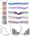
Figure 1
Figure 1
Heated metal rod burns heal through contraction and re-epithelialization. (a) Photographs and (b) images of Martius, Scarlet and Blue (MSB)-stained sections of healing skin at the indicated day post thermal burn. Scale is as indicated. (c) Burn closure is shown as a change in the percentage of the original burn area over-time. (d) Burn contraction is shown as the change in burn width over-time. (e) Burn re-epithelialization is shown as the change in the percentage neo-epidermal coverage over-time. Data represents the mean ± SEM, n = 8.
Figure 2
Figure 2
Application of a heated metal rod results in a partial-thickness skin burn. (a) Images of healing skin sections with terminal deoxynucleotidyl transferase-mediated deoxyuridine triphosphate nick-end labelling (TUNEL) at the indicated day post thermal burn. Scale is as indicated. (b) Burn depth over time is shown relative to the depth of full-thickness skin and the panniculus carnosus. Data represents the mean ± SEM, n = 6.
Figure 3
Figure 3
Heated metal rod burns result in a persistent scar. (a) Photographs and images of (b) MSB-stained and (c) picrosirius red-stained sections of skin scars at the indicated day post-thermal burn. Collagen deposition within the scars is shown at higher magnification in the right panel. Scale is as indicated. (d) Scar area is shown as a change in the percentage of the area over-time. (e) Epidermal scar index is shown as the change in epidermal width within the scar relative to adjacent skin over-time. (f) Dermal scar index is shown as the change in the dermal scar area relative to the average scar width over-time. (g) Collagen density is shown as the change in the percentage of the area of collagen staining over-time. Data represents the mean ± SEM, n = 8.
Figure 4
Figure 4
Profile of inflammatory cell populations during healing and scarring of heated rod burns. Images of skin sections stained with 4′,6-diamidino-2-phenylindole (DAPI) and antibodies against (a) Gr-1 and F4/80, (b) calprotectin and iNOS, (c) MHC II and CD207, or with (d) toluidine blue at the indicated day post thermal burn. Cell-specific staining is shown at higher magnification in the right panel. Scale is as indicated. Changes in the number of (e) Gr-1+ neutrophils, (f) F4/80+ macrophages, (g) calprotectin+ and iNOS+ macrophages, (h) MHC II+ DC, (i) CD207+ LC, and (j) toluidine blue+ mast cells within the burn/scar over time are expressed relative to area. Values for normal skin are as indicated. Data represents the mean ± SEM, n = 6.
Figure 5
Figure 5
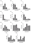
Profile of gene expression during healing and scarring of heated rod burns. Quantitative PCR was used to measure expression of (a) IL-1β, (b) IL-6, (c) TNF, (d) MCP-1, (e) MIP-2α, (f) IL-10, (g) VEGF-A, (h) TGF-β1, (i) TGF-β3 (j) Col3α1, and (k) Col1α2 in samples taken of healing skin. The level of mRNA is relative to that of TATA box binding protein (TBP) and normal skin. Data represents the mean ± SEM, n = 4.

Similar articles

Cited by

References

    1. WHO Fact Sheet: Burns. [(accessed on 6 March 2018)]; Available online: https://www.who.int/news-room/fact-sheets/detail/burns.
    1. Wang Y., Beekman J., Hew J., Jackson S., Issler-Fisher A.C., Parungao R., Lajevardi S.S., Li Z., Maitz P.K.M. Burn injury: Challenges and advances in burn wound healing, infection, pain and scarring. Adv. Drug Deliv. Rev. 2018;123:3–17. doi: 10.1016/j.addr.2017.09.018. - DOI - PubMed
    1. Monstrey S., Hoeksema H., Verbelen J., Pirayesh A., Blondeel P. Assessment of burn depth and burn wound healing potential. Burns. 2008;34:761–769. doi: 10.1016/j.burns.2008.01.009. - DOI - PubMed
    1. Khan A.A., Rawlins J., Shenton A.F., Sharpe D.T. The Bradford Burn Study: The epidemiology of burns presenting to an inner city emergency department. Emerg. Med. J. 2007;24:564–566. doi: 10.1136/emj.2005.027730. - DOI - PMC - PubMed
    1. Evers L.H., Bhavsar D., Mailander P. The biology of burn injury. Exp. Dermatol. 2010;19:777–783. doi: 10.1111/j.1600-0625.2010.01105.x. - DOI - PubMed