Review
. 2019 Jan 28;20(3):559.
doi: 10.3390/ijms20030559.

GPR68: An Emerging Drug Target in Cancer

Affiliations
Review

GPR68: An Emerging Drug Target in Cancer

Shu Z Wiley et al. Int J Mol Sci. .

Abstract

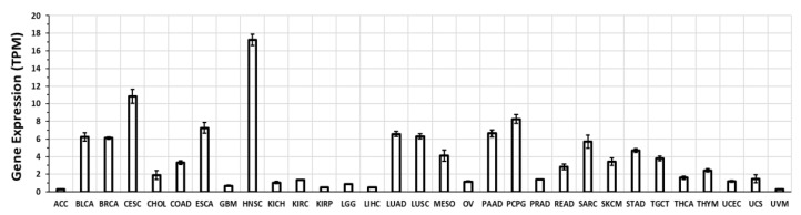
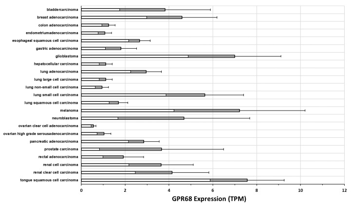
GPR68 (or ovarian cancer G protein-coupled receptor 1, OGR1) is a proton-sensing G-protein-coupled receptor (GPCR) that responds to extracellular acidity and regulates a variety of cellular functions. Acidosis is considered a defining hallmark of the tumor microenvironment (TME). GPR68 expression is highly upregulated in numerous types of cancer. Emerging evidence has revealed that GPR68 may play crucial roles in tumor biology, including tumorigenesis, tumor growth, and metastasis. This review summarizes current knowledge regarding GPR68-its expression, regulation, signaling pathways, physiological roles, and functions it regulates in human cancers (including prostate, colon and pancreatic cancer, melanoma, medulloblastoma, and myelodysplastic syndrome). The findings provide evidence for GPR68 as a potentially novel therapeutic target but in addition, we note challenges in developing drugs that target GPR68.

Keywords: GPR68; RNA-seq; acidosis; proton-sensing GPCRs; tumor microenvironment.


Conflict of interest statement

The authors declare no conflict of interest.

Figures

Figure 1
Figure 1
Immunoblotting of GPR68. (A) HEK293 cells were transfected with GPR68-v5tag plasmid (0–4 µg). After 48 h, cell lysates were prepared for immunoblotting using V5 antibody (#R960-25, Invitrogen). Three bands were observed, at 58, 41, and 38 kDa. (B) Immunoblotting of primary human pancreatic cancer associated fibroblasts (CAFs 1–5), pancreatic fibroblasts (PFs), and pancreatic stellate cells (PSCs) detected GPR68 at 58 kDa. (C) PF, PSC, and CAF samples treated with PNGase F (for deglycosylation) shifted the GPR68 band from 58 kDa to 41 kDa.
Figure 2
Figure 2
GPR68 expression in human tissues. These data were generated by RNA-seq, quantified in transcripts per million (TPM) from the GTEx database [33], re-analyzed via TOIL [34] and obtained from xena.ucsc.edu. The data shown are average +/− standard error of the mean (SEM) expression in each tissue type.
Figure 3
Figure 3
GPR68 expression in solid tumors. These data were generated by RNA-seq, quantified in transcripts per million (TPM) from the Cancer Genome Atlas (TCGA) database [64], re-analyzed via TOIL [34], and obtained from xena.ucsd.edu. Expression indicated are mean and standard error of the mean (SEM) in each tumor type. ACC (adrenocortical carcinoma); BLCA (bladder urothelial carcinoma); BRCA (breast invasive carcinoma); CESC (cervical squamous cell carcinoma and endocervical adenocarcinoma); CHOL (cholangiocarcinoma); COAD (colon adenocarcinoma); ESCA (esophageal carcinoma); GBM (glioblastoma multiforme); HNSC (head and neck squamous cell carcinoma); KICH (kidney chromophobe); KIRC (kidney renal clear cell carcinoma); KIRP (kidney renal papillary cell carcinoma); LGG (brain lower grade glioma); LIHC (liver hepatocellular carcinoma); LUAD (lung adenocarcinoma); LUSC (lung squamous cell carcinoma); MESO (mesothelioma); OV (ovarian serous cystadenocarcinoma); PAAD (pancreatic adenocarcinoma); PCPG (pheochromocytoma and paraganglioma); PRAD (prostate adenocarcinoma); READ (rectum adenocarcinoma); SARC (sarcoma); SKCM (skin cutaneous melanoma); STAD (stomach adenocarcinoma); TGCT (testicular germ cell tumors); THCA (thyroid carcinoma); THYM (thymoma); UCEC (uterine corpus endometrial carcinoma); UCS (uterine carcinosarcoma); UVM (uveal melanoma).
Figure 4
Figure 4
Differential expression of GPR68 in tumors compared to normal tissue. The Cancer Genome Atlas (TCGA) [64] tumors were subdivided into 45 histological subtypes as described previously [24]. Closed squares indicate the fold-changes in GPR68 expression that are statistically significant (false discovery rate (FDR) < 0.05); open squares indicate non-significant fold-changes. Baseline expression in normal tissue is set as 1.
Figure 5
Figure 5
GPR68 expression in cancer cell lines from a range of tumor types. These data were generated by RNA-seq, quantified in transcripts per million (TPM) and analyzed via the iRAP pipeline [66], from the Cancer Cell Line Encyclopedia [65] accessed via the EMBL-EBI expression atlas. GPR68 expression is plotted as mean and standard error of the mean (SEM) across all cell lines.
Figure 6
Figure 6
GPR68 expression in hematopoietic cells. These data are from the Blueprint Consortium, hosted at the EMBL-EBI expression atlas, and show GPR68 expression in transcripts per million (TPM) in a variety of cell types.
Figure 7
Figure 7
Summary of GPR68 functions in cancer cells and other cells in the tumor microenvironment (TME). GPR68 is expressed on a variety of cells in the TME, each of which has responses to GPR68 activation, as indicated on the figure. * GPR68 has not as-yet been directly shown on tumor endothelial cells, but based on data for vascular endothelial cells are likely to be present in tumors.

Similar articles

Cited by

References

    1. Vassilatis D.K., Hohmann J.G., Zeng H., Li F., Ranchalis J.E., Mortrud M.T., Brown A., Rodriguez S.S., Weller J.R., Wright A.C., et al. The G Protein-Coupled Receptor Repertoires of Human and Mouse. Proc. Natl. Acad. Sci. USA. 2003;100:4903–4908. doi: 10.1073/pnas.0230374100. - DOI - PMC - PubMed
    1. Heldin C., Lu B., Evans R., Gutkind J.S. Signals and Receptors. Cold Spring Harb. Perspect. Biol. 2016;8:a005900. doi: 10.1101/cshperspect.a005900. - DOI - PMC - PubMed
    1. Lynch J.R., Wang J.Y. G Protein-Coupled Receptor Signaling in Stem Cells and Cancer. Int. J. Mol. Sci. 2016;17:707. doi: 10.3390/ijms17050707. - DOI - PMC - PubMed
    1. O’Hayre M., Degese M., Gutkind J. Novel Insights into G Protein and G Protein-Coupled Receptor Signaling in Cancer. Curr. Opin. Cell Biol. 2014;27:126–135. doi: 10.1016/j.ceb.2014.01.005. - DOI - PMC - PubMed
    1. Neves S.R., Ram P.T., Iyengar R. G Protein Pathways. Science. 2002;296:1636–1640. doi: 10.1126/science.1071550. - DOI - PubMed

MeSH terms

Substances