. 2019 Apr 1;151(4):505-517.
doi: 10.1085/jgp.201812195. Epub 2019 Jan 29.

Functional characterization of the alanine-serine-cysteine exchanger of Carnobacterium sp AT7

Affiliations

Functional characterization of the alanine-serine-cysteine exchanger of Carnobacterium sp AT7

Paola Bartoccioni et al. J Gen Physiol. .

Abstract

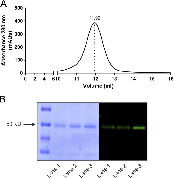
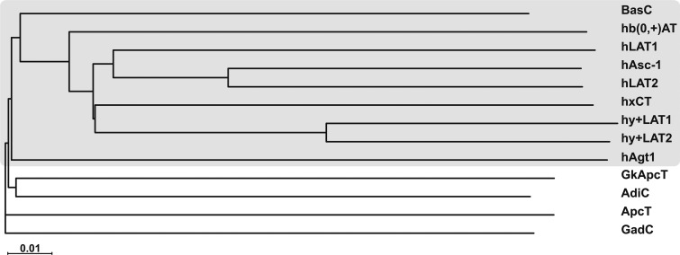
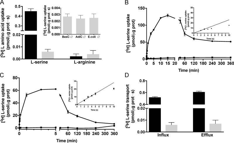
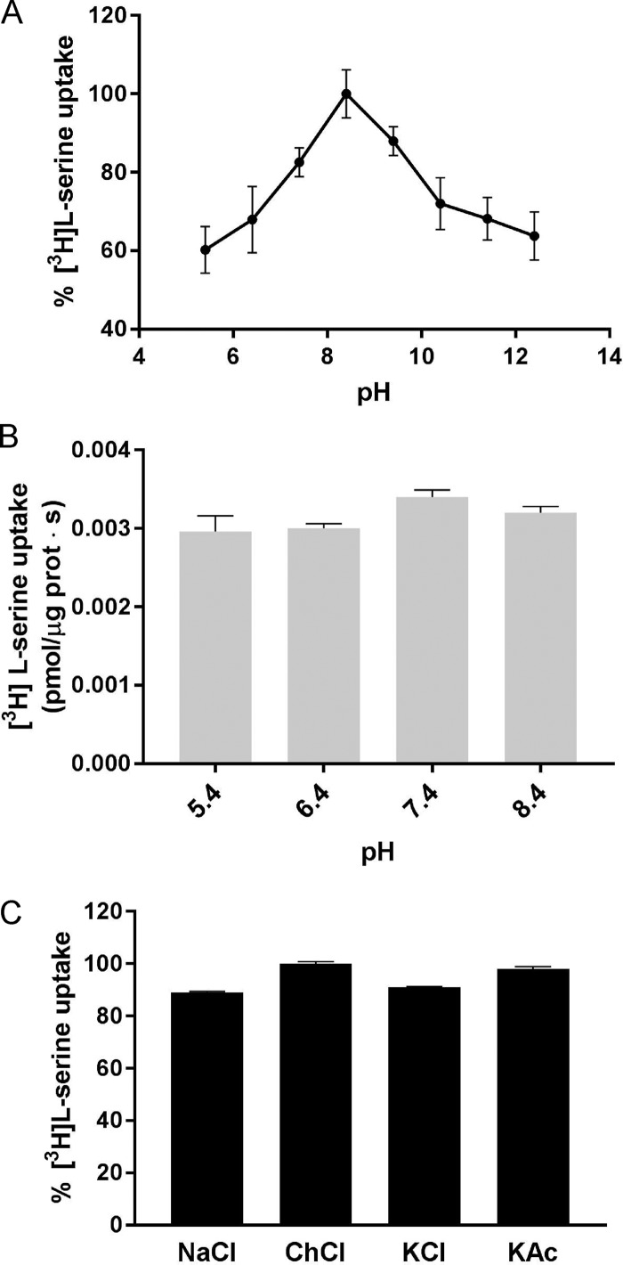
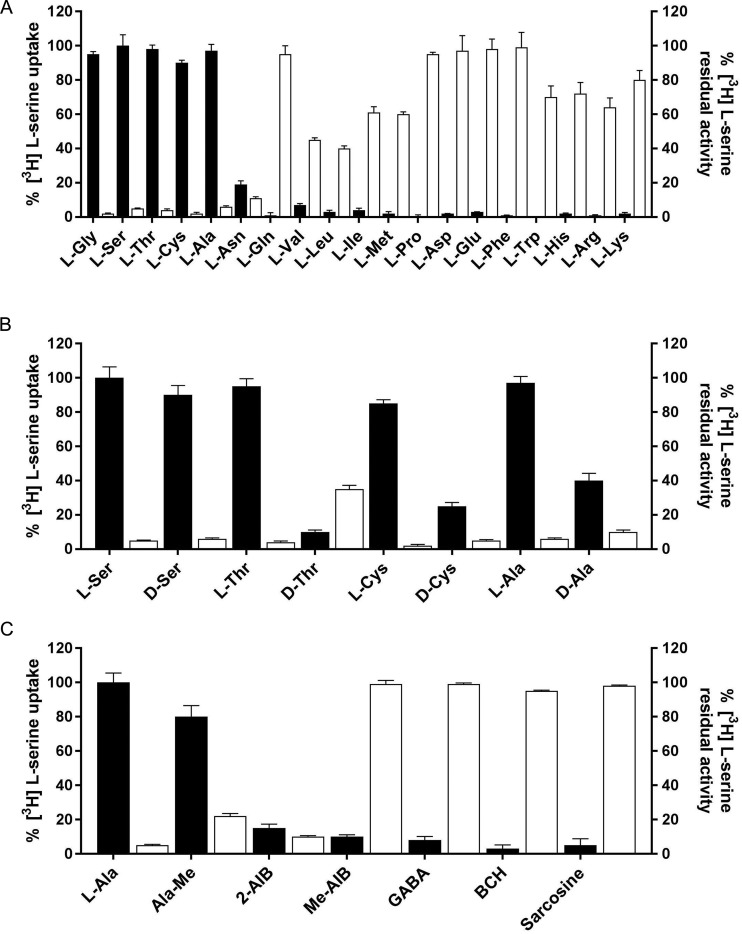
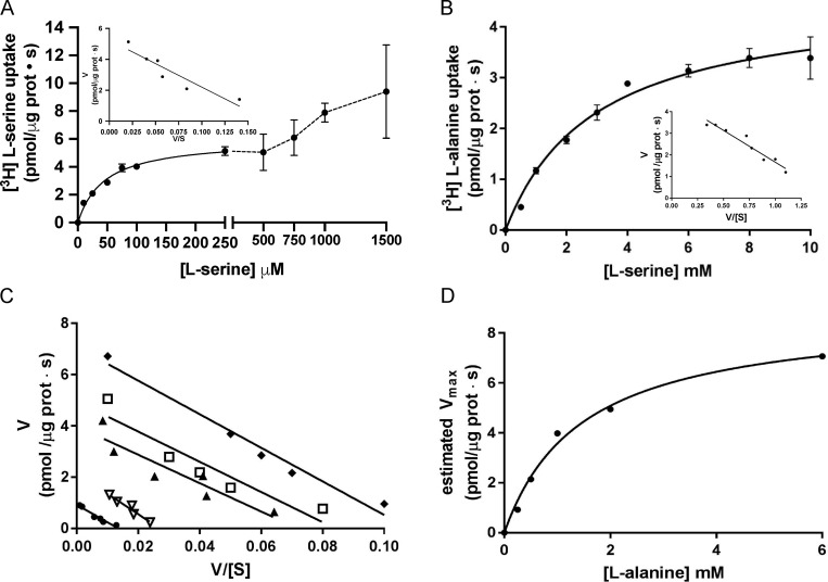
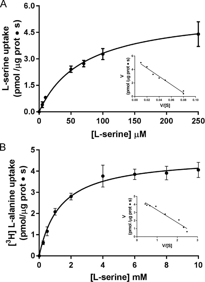
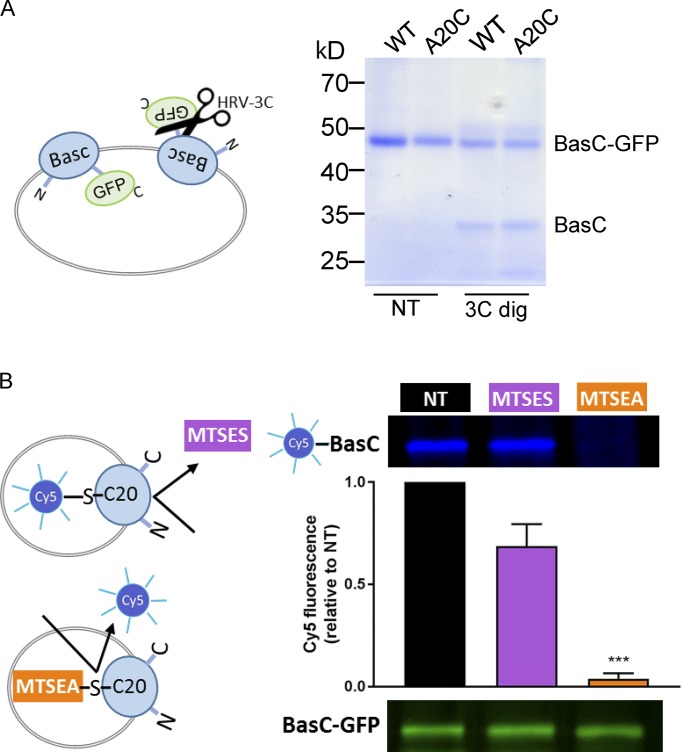
Many key cell processes require prior cell uptake of amino acids from the environment, which is facilitated by cell membrane amino acid transporters such as those of the L-type amino acid transporter (LAT) subfamily. Alterations in LAT subfamily amino acid transport are associated with several human diseases, including cancer, aminoacidurias, and neurodegenerative conditions. Therefore, from the perspective of human health, there is considerable interest in obtaining structural information about these transporter proteins. We recently solved the crystal structure of the first LAT transporter, the bacterial alanine-serine-cysteine exchanger of Carnobacterium sp AT7 (BasC). Here, we provide a complete functional characterization of detergent-purified, liposome-reconstituted BasC transporter to allow the extension of the structural insights into mechanistic understanding. BasC is a sodium- and proton-independent small neutral amino acid exchanger whose substrate and inhibitor selectivity are almost identical to those previously described for the human LAT subfamily member Asc-1. Additionally, we show that, like its human counterparts, this transporter has apparent affinity asymmetry for the intra- and extracellular substrate binding sites-a key feature in the physiological role played by these proteins. BasC is an excellent paradigm of human LAT transporters and will contribute to our understanding of the molecular mechanisms underlying substrate recognition and translocation at both sides of the plasma membrane.


Figures

Figure 1.
Figure 1.
BasC purification and reconstitution in PLs. (A) SEC of DDM-solubilized Ni2+-NTA purified BasC-GFP protein. mAUs, milli absorbance units. (B) Coomassie blue–stained gel and in-gel fluorescence of purified BasC. Purification of His-tagged BasC-GFP by nickel affinity chromatography with or without a further SEC step. Coomassie blue–stained (left) and in-gel fluorescence (right) SDS-PAGE (12% polyacrylamide gel) of the different purification steps is shown: Ni2+-NTA purified BasC-GFP (lane 1), BasC reconstituted in PLs (lane 2), and SEC-purified BasC (lane 3). BasC-GFP migrates as a prominent band at ∼50 kD.
Figure 2.
Figure 2.
Phylogenetic relationships of BasC with other prokaryotic and eukaryotic members of the APC family. The neighbor-joining tree shows the phylogenetic relationships of BasC with human members of the LAT subfamily, within the APC family, and the crystallized prokaryotic members of the APC family. The tree was performed using all the alignable member sequences within the APC family present in the Transporter Classification Database (www.tcdb.org).
Figure 3.
Figure 3.
Functional characterization of BasC reconstituted into E. coli liposomes. (A) 10 µM [3H]L-serine (1 µCi/µl) or [3H]L-arginine (1 µCi/µl) influx (pmol/µg protein · s) into BasC-GFP-PLs containing a mixture of 10 representative amino acids (L-Arg, L-Orn, Gly, L-Pro, L-Ala, L-Leu, L-Met, L-Phe, L-Tyr, and L-Glu at 1 mM each; black bars) or no amino acid (gray bars). Inset: 10 µM [3H]L-serine (1 µCi/µl) uptake (pmol/µg protein · s) in empty BasC-PLs, AdiC-PLs or E. coli polar lipid liposomes. Data (mean ± SEM) are from three experiments with three replicates per condition. (B) Time course (0–360 min) of 10 µM [3H]L-serine influx (pmol/µg protein) into BasC-GFP-PLs containing 4 mM L-serine (closed circles) or no amino acid (closed squares). Inset: Time course (0–10 s) of 10 µM [3H]L-serine/4 mM L-serine exchange (pmol/µg protein). Data correspond to a representative experiment, performed using three replicates. A second independent experiment gave similar results. (C) Time course (0–360 min) of 10 µM [3H]L-serine influx (pmol/µg protein) into BasC-GFP-PLs containing 4 mM L-alanine (closed circles) or no amino acid (closed squares). Inset: Time course (0–10 s) of 10 µM [3H]L-serine/4 mM L-alanine exchange (pmol/µg protein). Data correspond to a representative experiment, performed using three replicates. A second independent experiment gave similar results. (D) Comparison of 10 µM [3H]L-serine (1 µCi/data point) influx (left) and 10 µM [3H]L-serine (1 µCi/data point) efflux (right) in BasC-GFP-PLs, expressed in pmol/µg protein · s. Efflux measurements were performed by filling the liposomes by three freeze–thaw cycles with transport buffer plus 10 µM L-serine and 1 µCi/data point of [3H]L-serine. The release of [3H]L-serine was measured by adding 180 µl of transport buffer to the BasC-GFP-PL suspension with or without 4 mM cold amino acid. Efflux of radiolabeled L-serine was dramatically stimulated by the presence of L-serine in the external medium. Data (mean ± SEM) are from three experiments with three replicates per condition.
Figure 4.
Figure 4.
Effect of external pH and salts on BasC activity. (A) 10 µM [3H]L-serine (1 µCi/µl) influx into BasC-GFP-PLs containing 4 mM L-alanine at different pHs. Internal pH was maintained at 7.4 while external pH was changed (5.4–11.4). Transport was expressed as the percentage of [3H]L-serine transport, considering 100% the highest [3H]L-serine uptake value obtained. Data are from three experiments with three replicates per condition. (B) 10 µM [3H]L-serine (1 µCi/µl) influx (pmol/µg protein · s) into BasC-GFP-PLs containing no amino acid and at different pHs. Internal pH was maintained at 7.4 while external pH was changed (5.4–8.4). We observed [3H]L-serine exchange in all the assayed external proton concentrations, this exchange being dependent solely on the internal amino acid concentration. No significant increase in [3H]L-serine uptake was observed at any external pH value in empty PLs. Data (mean± SEM) are from three experiments with three replicates per condition. (C) Effect of ions on BasC activity. Uptake of 10 µM [3H]L-serine (1 µCi/µl) into BasC-PLs containing 4 mM L-alanine was performed in uptake buffer (20 mM Tris-Base and 150 mM NaCl, pH 7.4). NaCl was then replaced by 150 mM of either choline chloride (ChoCl), potassium chloride (KCl), or potassium acetate (KAc), and radiolabeled serine uptake was measured. Transport was expressed as the percentage of transport in BasC-GFP-PLs containing 4 mM L-alanine in uptake buffer with NaCl. Data are from three experiments with three replicates per condition.
Figure 5.
Figure 5.
Substrate and inhibitor specificity of BasC transport activity. (A) BasC substrate specificity. 10 µM [3H]L-serine (1 µCi/µl) influx into BasC-GFP-PLs containing 4 mM of the indicated individual amino acids (black bars). Transport was expressed as the percentage of the transport in BasC-PLs containing 4 mM L-alanine. 10 µM [3H]L-serine (1 µCi/µl) influx into BasC-GFP-PLs measured in the presence of 5 mM of the indicated amino acids in the external medium (white bars). Inhibition was expressed as the percentage of the transport in BasC-GFP-PLs containing 4 mM L-alanine with no cis-inhibitors. Data are from three experiments with three replicates per condition. (B) BasC stereoselectivity. 10 µM [3H]L-serine (1 µCi/µl) influx into BasC-PLs containing 4 mM of the indicated individual L– or D–amino acids (black bars). Transport was expressed as the percentage of the transport in BasC-GFP-PLs containing 4 mM L-alanine. 10 µM [3H]L-serine (1 µCi/µl) influx into BasC-GFP-PLs measured in the presence of 4 mM of the indicated L– or D–amino acids in the external medium (white bars). Inhibition was expressed as the percentage of transport in BasC-GFP-PLs containing 4 mM L-alanine with no cis-inhibitors. Data are from three experiments with three replicates per condition. (C) BasC amino acid derivative specificity. 10 µM [3H]L-serine (1 µCi/µl) influx into BasC-GFP-PLs containing 4 mM of the indicated individual amino acid derivatives (black bars). Transport was expressed as the percentage of transport in BasC-GFP-PLs containing 4 mM L-alanine. 10 µM [3H]L-serine (1 µCi/µl) influx into BasC-GFP-PLs measured in the presence of 4 mM of the indicated amino acid derivatives in the external medium (white bars). Inhibition was expressed as the percentage of transport in BasC-GFP-PLs containing 4 mM L-alanine with no cis-inhibitors. Data are from three experiments with three replicates per condition.
Figure 6.
Figure 6.
Kinetic characterization of [3H]L-serine uptake in BasC PLs. (A) Determination of extraliposomal kinetic parameters. Michaelis–Menten plot of the transporter-mediated uptake of [3H]L-serine (1 µCi/µl, 4 s pmol/µg protein · s) in BasC-GFP-PLs containing 4 mM L-alanine, varying extracellular L-serine concentrations (0–1,500 µM). Mediated transport (exchange) was calculated as [3H]L-serine uptake in PLs containing 4 mM L-alanine minus empty PLs. Data correspond to a representative experiment, performed using three replicates. Inset: Eadie–Hofstee plot of the kinetics covering only L-serine concentrations between 0–250 µM (i.e., the high apparent affinity component). Km and Vmax values were 45 ± 5 µM and 6.0 ± 0.2 pmol [3H]L-serine/µg prot · s, respectively. V/[S]: pmol [3H]L-serine/µg prot · s / substrate concentration. (B) Determination of intraliposomal kinetic parameters. Michaelis–Menten plot of the transporter-mediated uptake of [3H]L-alanine (10 µM, 1 µCi/µl, pmol/µg protein · s) in BasC-GFP-PLs containing 0.5–10 mM cold L-serine. Mediated transport was calculated as [3H]L-alanine uptake in L-serine–containing PLs minus empty PLs. Data correspond to a representative experiment, performed using three replicates. Inset: Eadie–Hofstee plot. Km value was 2.5 ± 0.4 mM. In A and B, three independent experiments were performed, giving similar results. (C) Extraliposomal kinetics as in A at different intraliposomal concentrations of L-alanine (0.2, 0.5, 1.0, 2.0, and 6.0 mM). Eadie–Hofstee plots from a representative experiment with the mean values from three replicates. (D) Michaelis–Menten plot of the estimated Vmax values at the different intraliposomal L-alanine concentrations from the kinetic series shown in C.
Figure 7.
Figure 7.
Extraliposomal and intraliposomal kinetics of BasC without GFP. (A) Determination of extraliposomal kinetic parameters. Michaelis–Menten plot of the transporter-mediated uptake of [3H]L-serine (1 µCi, 4 s) in BasC-PLs containing 4 mM L-alanine, varying extracellular L-serine concentrations (0–250 µM). Mediated transport was calculated as [3H]L-serine uptake in PLs containing 4 mM L-alanine minus empty PLs. Inset: Eadie–Hofstee plot. Km and Vmax values were 71.7 ± 4.3 µM and 5.7 ± 0.1 pmol [3H]L-serine/µg prot · s, respectively. (B) Determination of intraliposomal kinetic parameters. Michaelis–Menten plot of the transporter-mediated uptake of [3H]L-alanine (10 µM, 1 µCi, 4 s) in BasC-PLs containing 0.5–10 mM cold L-serine. Mediated transport was calculated as [3H]L-alanine uptake in L-serine–containing PLs minus empty PLs. In A and B, data correspond to a representative experiment, performed using three replicates. Inset: Eadie–Hofstee plot. Km value was 1.3 ± 0.1 mM. Data (mean ± SEM) correspond to triplicates from representative experiments (A and B).
Figure 8.
Figure 8.
Vectorial modification of BasC A20C mutant by MTSES from the extracellular side. (A) Random incorporation of the BasC-GFP protein in PLs. BasC-GFP-PLs for wild-type and A20C mutant were treated with HRV-3C protease (3C dig) for 2 h at 4°C. The protease site is accessible in the inside-out BasC-GFP–inserted molecules, resulting in GFP cleavage. Protease-treated PLs were then mixed with loading sample buffer and analyzed in a 10% polyacrylamide gel. As a result, approximately half of the wild-type and mutant proteins were inserted right-side-out when reconstituted in E. coli polar lipid liposomes. (B) MTSEA but not MTSES pretreatment of BasC-A20C-GFP-PLs protected inside-out molecules from Cy5-maleimide dyeing. BasC-A20C-GFP-PLs were digested with HRV-3C protease for 2 h at 4°C to remove GFP from the inside-out BasC-A20C molecules. Then, either MTSES (1 mM; 15 min) or MTSEA (5 mM; 30 min) pretreatment was performed before BasC-A20C labeling with the membrane-permeable reagent Cy5-maleimide (1 mM; overnight). Finally, samples were mixed with loading sample buffer and analyzed in a 10% polyacrylamide gel. In-gel fluorescence at 600 nm (GFP) and 700 nm (Cy5) is shown. The graph represents quantification of Cy5 fluorescence in BasC-A20C normalized by GFP fluorescence in BasC-A20C-GFP and relative to aliquots nontreated with MTSES or MTSES in each experiment (NT). Data (mean ± SEM) are from four independent experiments. Student’s t test for paired data was used for statistical comparison between NT and MTSES (no significant differences) or MTSEA (***, P ≤ 0.001) treatments.
Figure 9.
Figure 9.
Sidedness of the transport activity of BasC in PLs. (A and B) Effect of cysteine-modifying reagents MTSES (ES) and MTSEA (EA) on L-serine transport activity. Time course (0–60 s) influx of 10 µM [3H]L-serine (0.5 µCi/data point) in empty (empty circles) or filled with 4 mM L-alanine (filled circles) BasC-A20C-GFP-PLs (A) and time course (0–60 s) efflux of 10 µM [3H]L-serine (0.5 µCi/data point) filled BasC-A20C-GFP-PLs against medium without amino acid (empty circles) or with 4 mM alanine and nontreated (filled circles), treated with 1 mM MTSES (ES) for 15 min (magenta circles), or treated with 5 mM MTSEA (EA) for 30 min (orange circles; B). 10 µM [3H]L-serine influx and efflux occurs via right-side-out– and inside-out–oriented BasC in PLs, respectively. MTSES only reaches Cys 20 (C20) in right-side-out–oriented BasC. MTSEA reaches C20 independently of the orientation of BasC insertion in PLs. Influx was determined by the incorporation of [3H]L-serine into PLs, whereas efflux was determined by the decay of [3H]L-serine remaining in PLs. MTSES or MTSEA treatment abolished L-serine influx, while only treatment with MTSEA, but not MTSES, completely inhibited L-serine efflux. Data (mean ± SEM) correspond to quadruplicates of a representative experiment. Two additional independent experiments gave similar results. (C and D) Kinetics of [3H]L-serine/4 mM L-alanine exchange in BasC-A20C-GFP-PLs treated (magenta circles) and nontreated (black circles) with 1 mM MTSES for 15 min. (D) Eadie–Hofstee plot of the kinetics shown in C. Nontreated PLs (black circles) showed a complex kinetics with low (line) and high Km components. MTSES treatment abolished the low Km (i.e., apparent high-affinity) component. Data (mean ± SEM) correspond to quadruplicates of a representative experiment. Another independent experiment gave similar results.

References

    1. Altschul S.F., Gish W., Miller W., Myers E.W., and Lipman D.J.. 1990. Basic local alignment search tool. J. Mol. Biol. 215:403–410. 10.1016/S0022-2836(05)80360-2 - DOI - PubMed
    1. Borsani G., Bassi M.T., Sperandeo M.P., De Grandi A., Buoninconti A., Riboni M., Manzoni M., Incerti B., Pepe A., Andria G., et al. 1999. SLC7A7, encoding a putative permease-related protein, is mutated in patients with lysinuric protein intolerance. Nat. Genet. 21:297–301. 10.1038/6815 - DOI - PubMed
    1. Brindley A.A., Pickersgill R.W., Partridge J.C., Dunstan D.J., Hunt D.M., and Warren M.J.. 2008. Enzyme sequence and its relationship to hyperbaric stability of artificial and natural fish lactate dehydrogenases. PLoS One. 3:e2042 10.1371/journal.pone.0002042 - DOI - PMC - PubMed
    1. Brown J.M., Hunihan L., Prack M.M., Harden D.G., Bronson J., Dzierba C.D., Gentles R.G., Hendricson A., Krause R., Macor J.E., and Westphal R.S.. 2014. In vitro Characterization of a small molecule inhibitor of the alanine serine cysteine transporter -1 (SLC7A10). J. Neurochem. 129:275–283. 10.1111/jnc.12618 - DOI - PubMed
    1. Calonge M.J., Gasparini P., Chillarón J., Chillón M., Gallucci M., Rousaud F., Zelante L., Testar X., Dallapiccola B., Di Silverio F., et al. 1994. Cystinuria caused by mutations in rBAT, a gene involved in the transport of cystine. Nat. Genet. 6:420–425. 10.1038/ng0494-420 - DOI - PubMed

Publication types

MeSH terms

Substances