

Competition in Biofilms between Cystic Fibrosis Isolates of Pseudomonas aeruginosa Is Shaped by R-Pyocins
- PMID: 30696740
- PMCID: PMC6355985
- DOI: 10.1128/mBio.01828-18
Competition in Biofilms between Cystic Fibrosis Isolates of Pseudomonas aeruginosa Is Shaped by R-Pyocins
Abstract
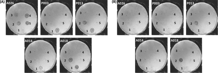
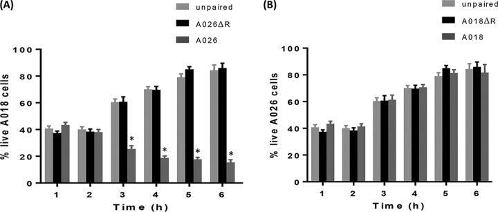
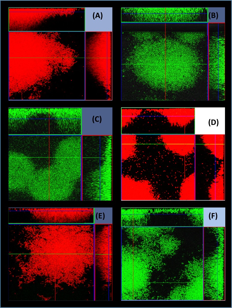
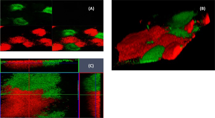
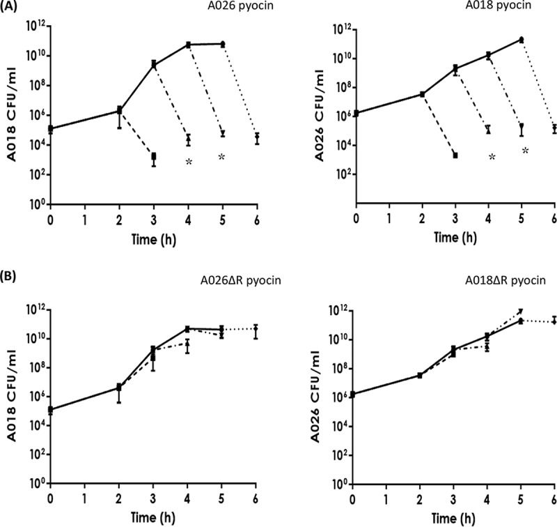
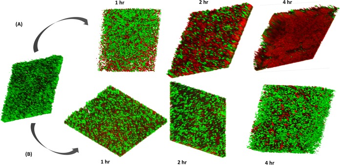
Pseudomonas aeruginosa is an opportunistic pathogen and the leading cause of morbidity and mortality in cystic fibrosis (CF) patients. P. aeruginosa infections are difficult to treat due to a number of antibiotic resistance mechanisms and the organism's propensity to form multicellular biofilms. Epidemic strains of P. aeruginosa often dominate within the lungs of individual CF patients, but how they achieve this is poorly understood. One way that strains of P. aeruginosa can compete is by producing chromosomally encoded bacteriocins, called pyocins. Three major classes of pyocin have been identified in P. aeruginosa: soluble pyocins (S types) and tailocins (R and F types). In this study, we investigated the distribution of S- and R-type pyocins in 24 clinical strains isolated from individual CF patients and then focused on understanding their roles in interstrain competition. We found that (i) each strain produced only one R-pyocin type, but the number of S-pyocins varied between strains, (ii) R-pyocins were generally important for strain dominance during competition assays in planktonic cultures and biofilm communities in strains with both disparate R- and S-pyocin subtypes, and (iii) purified R-pyocins demonstrated significant antimicrobial activity against established biofilms. Our work provides support for a role played by R-pyocins in the competition between P. aeruginosa strains and helps explain why certain strains and lineages of P. aeruginosa dominate and displace others during CF infection. Furthermore, we demonstrate the potential of exploiting R-pyocins for therapeutic gains in an era when antibiotic resistance is a global concern.IMPORTANCE A major clinical problem caused by Pseudomonas aeruginosa, is chronic biofilm infection of the lungs in individuals with cystic fibrosis (CF). Epidemic P. aeruginosa strains dominate and displace others during CF infection, but these intraspecies interactions remain poorly understood. Here we demonstrate that R-pyocins (bacteriocins) are important factors in driving competitive interactions in biofilms between P. aeruginosa strains isolated from different CF patients. In addition, we found that these phage-like pyocins are inhibitory against mature biofilms of susceptible strains. This highlights the potential of R-pyocins as antimicrobial and antibiofilm agents at a time when new antimicrobial therapies are desperately needed.
Keywords: Pseudomonas aeruginosa; bacteriocins; biofilms; cystic fibrosis; pyocins.
Copyright © 2019 Oluyombo et al.
Figures
References
-
- Rahme LG, Stevens EJ, Wolfort SF, Shao J, Tompkins RG, Ausubel FM. 1995. Common virulence factors for bacterial pathogenicity in plants and animals. Science 268:1899–1902. - PubMed
-
- Jorth P, Staudinger BJ, Wu X, Hisert KB, Hayden H, Garudathri J, Harding CL, Radey MC, Rezayat A, Bautista G, Berrington WR, Goddard AF, Zheng C, Angermeyer A, Brittnacher MJ, Kitzman J, Shendure J, Fligner CL, Mittler J, Aitken ML, Manoil C, Bruce JE, Yahr TL, Singh PK. 2015. Regional isolation drives bacterial diversification within cystic fibrosis lungs. Cell Host Microbe 18:307–319. doi: 10.1016/j.chom.2015.07.006. - DOI - PMC - PubMed
-
- Darch SE, McNally A, Harrison F, Corander J, Barr HL, Paszkiewicz K, Holden S, Fogarty A, Crusz SA, Diggle SP. 2015. Recombination is a key driver of genomic and phenotypic diversity in a Pseudomonas aeruginosa population during cystic fibrosis infection. Sci Rep 5:7649. doi: 10.1038/srep07649. - DOI - PMC - PubMed
Publication types
MeSH terms
Substances
LinkOut - more resources
Full Text Sources
Medical
