. 2019 Jan 29;9(1):826.
doi: 10.1038/s41598-018-36811-4.

Caspase-4 disaggregates lipopolysaccharide micelles via LPS-CARD interaction

Affiliations

Caspase-4 disaggregates lipopolysaccharide micelles via LPS-CARD interaction

Jinsu An et al. Sci Rep. .

Abstract

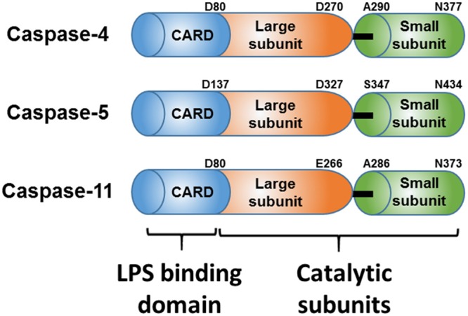
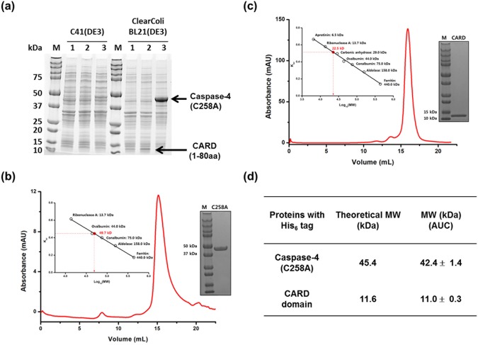
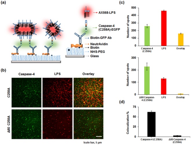
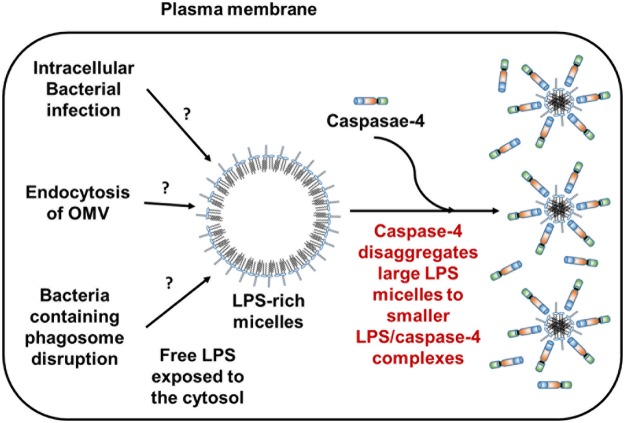
Lipopolysaccharides (LPS) are a major component of the outer membrane of Gram-negative bacteria and are pathogen-associated molecular patterns recognized by the TLR4/MD2 complex that induces an inflammatory response. Recently, the cytosolic receptors caspase-4/-5/-11 that bind LPS inside the cell and trigger inflammasome activation or pyroptosis, have been identified. Despite the important roles of caspase-4 in human immune responses, few studies have investigated its biochemical characteristics and interactions with LPS. Since caspase-4 (C258A) purified from an Escherichia coli host forms aggregates, monomeric proteins including full-length caspase-4, caspase-4 (C258A), and the CARD domain of caspase-4 have been purified from the insect cell system. Here, we report the overexpression and purification of monomeric caspase-4 (C258A) and CARD domain from E. coli and demonstrate that purified caspase-4 (C258A) and CARD domain bind large LPS micelles and disaggregate them to small complexes. As the molar ratio of caspase-4 to LPS increases, the size of the caspase-4/LPS complex decreases. Our results present a new function of caspase-4 and set the stage for structural and biochemical studies, and drug discovery targeting LPS/caspase-4 interactions by establishing a facile purification method to obtain large quantities of purified caspase-4 (C258A) and the CARD domain.


Conflict of interest statement

Korea Institute of Science and Technology has filed a patent on which H.S.C., E.G.Y., S.Y.K. and J.S.A. are listed as inventors. The other authors declare no competing interests.

Figures

Figure 1
Figure 1
Schematic domain structures of the cytosolic LPS receptors Caspase-4, Caspase-5 and Caspase-11. The amino acid numbering above corresponds to that in the UniProt database.
Figure 2
Figure 2
Monomeric caspase-4(C258A) and CARD purified from E. coli (a) A 4–20% SDS-PAGE gel comparing the expression levels of recombinant proteins in two different E. coli hosts. Whole cell lysates from pET28b empty vector (Lane 1), pET28b harboring the CARD domain of caspase-4 (Lane 2), and pET28b harboring caspase-4 (C258A) (Lane 3). The amount of protein loaded on the SDS gel was normalized to a cell culture biomass based on OD600. SEC elution profiles of caspase-4 (C258A) (b) and CARD domain (c) from Superdex 200 10/300 gl columns calibrated with protein standards and SDS-PAGE gels of the purified caspase-4 (C258A) (b) and CARD domain (c). 5 µg of purified protein was loaded in each lane. Calibration curves are presented in insets and enlarged calibration curves are presented in Supplementary Fig. S5. (d) Molecular weight (MW) determination of purified caspase-4 (C258A) and CARD domain from ClearColi BL21(DE3) by analytical ultracentrifugation (AUC) and theoretical MW. Shown are the mean values ± standard deviation. SDS-PAGE gels shown in a, b, and c were cropped for clarity and full-length gels are presented in Supplementary Figs S1 and S4.
Figure 3
Figure 3
E. coli purified caspase-4(C258A) and CARD form LPS/protein complexes in a molar ratio dependent manner. SEC elution profiles of different molar ratios of Ra-LPS/Caspase-4 (C258A) (a), Ra-LPS/Δ80 caspase-4 (C258A) (b), and Ra-LPS/CARD domain complex (c) from Superdex200 10/300 gl columns calibrated with protein standards. Calibration curves are presented in insets and enlarged calibration curves are presented in Supplementary Fig. S6.
Figure 4
Figure 4
Caspase-4 disaggregates large LPS micelles to smaller LPS/protein complexes via the CARD domain. TEM images of different molar ratios of Ra-LPS/caspase-4 (C258A) (a), Ra-LPS/Δ80 caspase-4 (C258A) (b), Ra-LPS/CARD (c), and Ra-LPS/LBP (d) were visualized by negative-staining. Scale bar represents 50 nm.
Figure 5
Figure 5
Single-molecule co-localization between EGFP tagged caspase-4 (C258A) (or Δ80 caspase-4 (C258A)) and AX568-LPS. Caspase-4 (C258A)-EGFP (or Δ80 caspase-4 (C258A)) and AX568-LPS were imaged after immobilization of 100 pM of Caspase-4 with anti-GFP followed by incubation with 6 µM LPS in the presence of 1 µM of AX568-LPS. (a) Schematic of the single-molecule co-localization assay and surface modification. (b) Upper panel: Representative images (25 × 25 µm) for caspase-4 (C258A)-EGFP (left) and AX568-LPS (middle), and their overlapped images (right). Lower panel: Representative images (25 × 25 µm) for Δ80 caspase-4 (C258A)-EGFP (left) and AX568-LPS (middle), and their overlapped images (right). Scale bar, 5 µm. (c) Average number of fluorescent spots for caspase-4 (green), LPS (red), and co-localized caspase-4 (C258A) (or Δ80 caspase-4 (C258A)) and LPS (orange) per image. ± indicates standard error of mean (SEM). (d) Average co-localization efficiency was determined by calculating the number of co-localized spots divided by the number of caspase-4 (C258A) (or Δ80 caspase-4 (C258A)) spots.
Figure 6
Figure 6
Proposed role of caspase-4 in response to LPS-rich micelles exposed to cytosol. Caspase-4 not only binds LPS-rich micelles but also disaggregates them to smaller LPS/caspase-4 complexes.

References

    1. Raetz CR, Whitfield C. Lipopolysaccharide endotoxins. Annual review of biochemistry. 2002;71:635–700. doi: 10.1146/annurev.biochem.71.110601.135414. - DOI - PMC - PubMed
    1. Tobias PS, Soldau K, Gegner JA, Mintz D, Ulevitch RJ. Lipopolysaccharide binding protein-mediated complexation of lipopolysaccharide with soluble CD14. J Biol Chem. 1995;270:10482–10488. doi: 10.1074/jbc.270.18.10482. - DOI - PubMed
    1. Park BS, et al. The structural basis of lipopolysaccharide recognition by the TLR4–MD-2 complex. nature. 2009;458:1191–1195. doi: 10.1038/nature07830. - DOI - PubMed
    1. Ryu J-K, et al. Reconstruction of LPS Transfer Cascade Reveals Structural Determinants within LBP, CD14, and TLR4-MD2 for Efficient LPS Recognition and Transfer. Immunity. 2017;46:38–50. doi: 10.1016/j.immuni.2016.11.007. - DOI - PubMed
    1. Poltorak A, et al. Defective LPS signaling in C3H/HeJ and C57BL/10ScCr mice: mutations in Tlr4 gene. Science. 1998;282:2085–2088. doi: 10.1126/science.282.5396.2085. - DOI - PubMed

Publication types