. 2019 Jan 29;9(1):900.
doi: 10.1038/s41598-018-36954-4.

Nanoparticle administration method in cell culture alters particle-cell interaction

Affiliations

Nanoparticle administration method in cell culture alters particle-cell interaction

Thomas L Moore et al. Sci Rep. .

Abstract

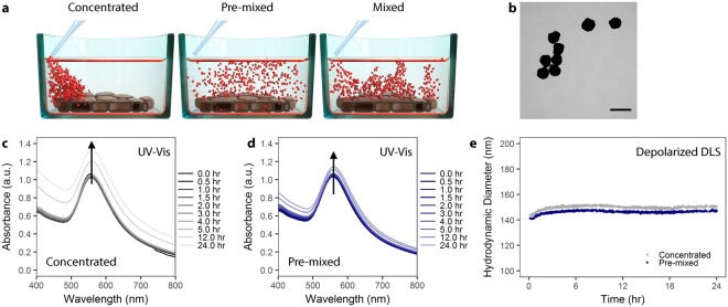
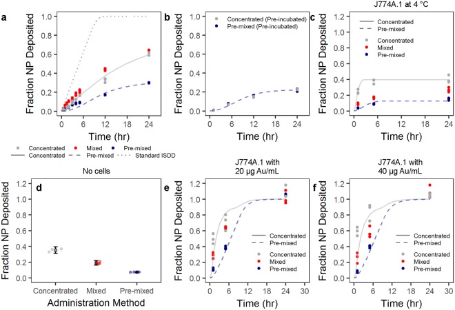
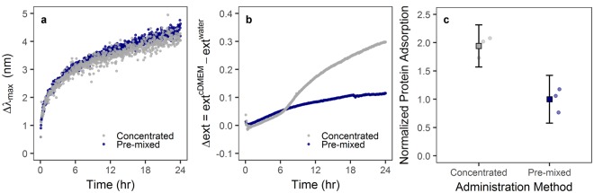
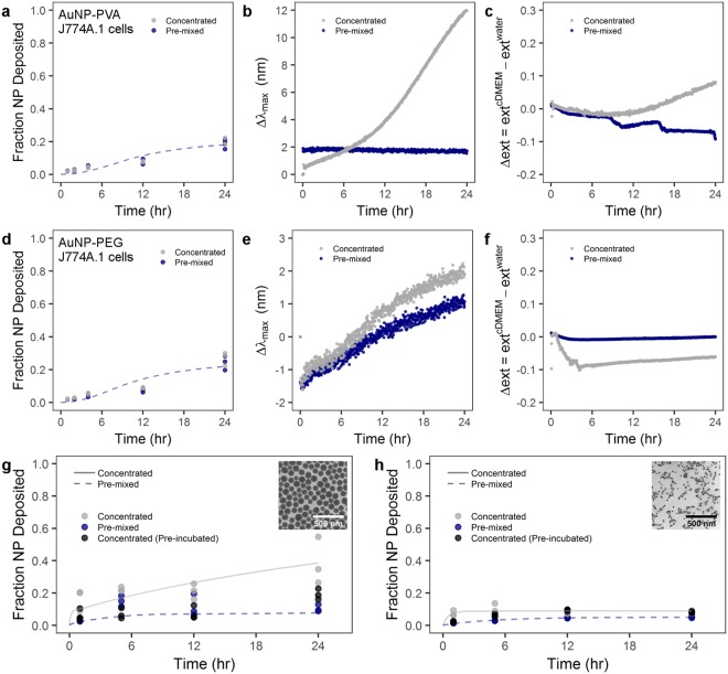
As a highly interdisciplinary field, working with nanoparticles in a biomedical context requires a robust understanding of soft matter physics, colloidal behaviors, nano-characterization methods, biology, and bio-nano interactions. When reporting results, it can be easy to overlook simple, seemingly trivial experimental details. In this context, we set out to understand how in vitro technique, specifically the way we administer particles in 2D culture, can influence experimental outcomes. Gold nanoparticles coated with poly(vinylpyrrolidone) were added to J774A.1 mouse monocyte/macrophage cultures as either a concentrated bolus, a bolus then mixed via aspiration, or pre-mixed in cell culture media. Particle-cell interaction was monitored via inductively coupled plasma-optical emission spectroscopy and we found that particles administered in a concentrated dose interacted more with cells compared to the pre-mixed administration method. Spectroscopy studies reveal that the initial formation of the protein corona upon introduction to cell culture media may be responsible for the differences in particle-cell interaction. Modeling of particle deposition using the in vitro sedimentation, diffusion and dosimetry model helped to clarify what particle phenomena may be occurring at the cellular interface. We found that particle administration method in vitro has an effect on particle-cell interactions (i.e. cellular adsorption and uptake). Initial introduction of particles in to complex biological media has a lasting effect on the formation of the protein corona, which in turn mediates particle-cell interaction. It is of note that a minor detail, the way in which we administer particles in cell culture, can have a significant effect on what we observe regarding particle interactions in vitro.


Conflict of interest statement

The authors declare no competing interests.

Figures

Figure 1
Figure 1
AuNP colloidal stability is independent of administration method. (a) Schematic showing the different administration method concepts. Particles can be administered to cells as a concentrated dose (left), a pre-mixed solution (center), or as a concentrated dose which is mixed via aspiration (right). (b) Transmission electron microscopy of citrate AuNP with shows a mean core diameter of 116 ± 12 nm (n = 289). Scale bar represents 200 nm. (c) UV-Vis spectra of PVP-coated AuNPs over 24 hr in cell culture media supplemented with 10% fetal bovine serum (FBS) following concentrated administration. (d) UV-Vis spectra of PVP-coated AuNPs administered via the pre-mixed method in cell culture media supplemented with 10% FBS. (e) Depolarized dynamic light scattering of PVP-coated AuNPs measured over 24 hr in complete cell culture media.
Figure 2
Figure 2
The deposition/uptake of AuNP-PVP (yellow) on J774A.1 cells was showed by overlaying enhanced darkfield microscopy images (AuNPs pseudo-colored yellow) onto fluorescent microscopy images of cells stained for actin (cyan) and cell nuclei (magenta). Scale bars represent 10 μm.
Figure 3
Figure 3
Particle cell interaction is dependent on administration method. (a) Particles administered as a concentrated, mixed, or pre-mixed dose showed variable uptake by J774A.1 mouse monocyte/macrophages as determined by ICP-OES. (b) This phenomenon was mitigated by first incubating the particle at a higher concentration in complete cell culture media, and then either adding as a concentrated dose or pre-mixing (pre-incubation). The trend in administration-dependent cellular interaction was conserved over various scenarios. (c) Administration at 4 °C was conducted to reduce active cellular uptake. (d) Non-specific NP adsorption to the culture plate was tested by incubating particles in complete cell culture medium overnight. Circles represent single data points and boxes represent mean ± one standard deviation. (e,f) AuNP-PVP deposition at 20 and 40 μg Au/mL, respectively, was tested over 24 hr. Lines indicate modified ISDD fit of data with concentrated or pre-mixed assumptions, as well as the standard ISDD conditions.
Figure 4
Figure 4
Particle introduction into complete cell culture media mediates protein adsorption to the particle surface. (a) UV-Vis analysis of AuNP-PVP over 24 hr showed a change in peak intensity wavelength. (b) UV-Vis analysis further showed a change in extinction intensity (Δext) dependent on administration method. These effects are indicative of protein adsorption to the nanoparticle surface. (c) Analysis of protein adsorption showed that, after 24 hr incubation in complete cell culture medium, AuNP-PVP administered via the concentrated route had an approximately 2-fold increase in protein adsorption compared to nanoparticles administered via the pre-mixed route.
Figure 5
Figure 5
Administration method effect on particle-cell interaction is heavily dependent on particle material, size, and surface coating. Cellular uptake by J774A.1 murine cells and UV-Vis analysis of (ac) PVA-coated AuNPs and (df) PEG-coated AuNPs. J774A.1 uptake of (g) PVP-coated SiO2 NPs and (h) PVP-coated superparamagnetic iron oxide NPs. (b,e) LSPR band shift for PVA-coated and PEG-coated AuNPs, respectively, following the concentrated or pre-mixed administration. (c,f) Time-dependent change of the extinction intensity (Δext) with respect to the extinction in water at maximum for PVA-coated and PEG-coated AuNP, respectively. Lines indicate modified ISDD fit of data with concentrated or pre-mixed assumptions. Inset TEM micrographs show (g) PVP-coated SiO2 and (h) PVP-coated superparamagnetic iron oxide NPs. Scale bars represent 500 nm.

References

    1. Kamaly N, Xiao Z, Valencia PM, Radovic-Moreno AF, Farokhzad OC. Targeted polymeric therapeutic nanoparticles: design, development and clinical translation. Chem. Soc. Rev. 2012;41:2971–3010. doi: 10.1039/c2cs15344k. - DOI - PMC - PubMed
    1. Venditto VJ, Szoka FC. Cancer nanomedicines: So many papers and so few drugs! Adv. Drug Deliv. Rev. 2013;65:80–88. doi: 10.1016/j.addr.2012.09.038. - DOI - PMC - PubMed
    1. Anselmo AC, Mitragotri S. An overview of clinical and commercial impact of drug delivery systems. J. Control. Rel. 2014;190:15–28. doi: 10.1016/j.jconrel.2014.03.053. - DOI - PMC - PubMed
    1. Tinkle S, et al. Nanomedicines: addressing the scientific and regulatory gap. Ann. N.Y. Acad. Sci. 2014;1313:35–56. doi: 10.1111/nyas.12403. - DOI - PubMed
    1. Min Y, Caster JM, Eblan MJ, Wang AZ. Clinical translation of nanomedicine. Chem. Rev. 2015;115:11147–11190. doi: 10.1021/acs.chemrev.5b00116. - DOI - PMC - PubMed

Publication types