Hyperautofluorescent Dots are Characteristic in Ceramide Kinase Like-associated Retinal Degeneration
- PMID: 30696906
- PMCID: PMC6351646
- DOI: 10.1038/s41598-018-37578-4
Hyperautofluorescent Dots are Characteristic in Ceramide Kinase Like-associated Retinal Degeneration
Abstract
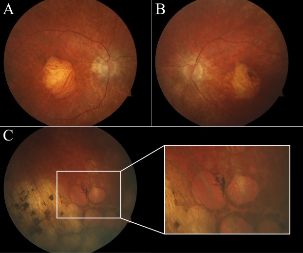
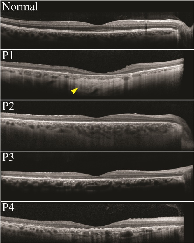
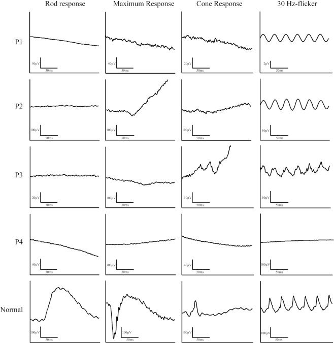
There is a lack of studies which seek to discern disease expression in patients with mutations that alter retinal ceramide metabolism, specifically in the ceramide kinase like (CERKL) gene. This cross-sectional case series reports a novel phenotypic manifestation of CERKL-associated retinopathy. Four unrelated patients with homozygous CERKL mutations underwent a complete ocular exam, spectral-domain optical coherence tomography, short-wavelength fundus autofluorescence (SW-AF), quantitative autofluorescence (qAF), and full-field electroretinogram (ffERG). Decreased visual acuity and early-onset maculopathy were present in all patients. All four patients had extensive hyperautofluorescent foci surrounding an area of central atrophy on SW-AF imaging, which has not been previously characterized. An abnormal spatial distribution of qAF signal was seen in one patient, and abnormally elevated qAF8 signal in another patient. FfERG recordings showed markedly attenuated rod and cone response in all patients. We conclude that these patients exhibit several features that, collectively, may warrant screening of CERKL as a first candidate: early-onset maculopathy, severe generalized retinal dysfunction, peripheral lacunae, intraretinal pigment migration, and hyperautofluorescent foci on SW-AF.
Conflict of interest statement
The authors declare no competing interests.
Figures


Similar articles
-
A New Cerkl Mouse Model Generated by CRISPR-Cas9 Shows Progressive Retinal Degeneration and Altered Morphological and Electrophysiological Phenotype.Invest Ophthalmol Vis Sci. 2020 Jul 1;61(8):14. doi: 10.1167/iovs.61.8.14. Invest Ophthalmol Vis Sci. 2020. PMID: 32658961 Free PMC article.
-
Primary versus Secondary Elevations in Fundus Autofluorescence.Int J Mol Sci. 2023 Aug 2;24(15):12327. doi: 10.3390/ijms241512327. Int J Mol Sci. 2023. PMID: 37569703 Free PMC article. Review.
-
Quantitative Fundus Autofluorescence and Genetic Associations in Macular, Cone, and Cone-Rod Dystrophies.Ophthalmol Retina. 2020 Jul;4(7):737-749. doi: 10.1016/j.oret.2020.02.009. Epub 2020 Feb 27. Ophthalmol Retina. 2020. PMID: 32646556
-
CERKL mutations cause an autosomal recessive cone-rod dystrophy with inner retinopathy.Invest Ophthalmol Vis Sci. 2009 Dec;50(12):5944-54. doi: 10.1167/iovs.09-3982. Epub 2009 Jul 2. Invest Ophthalmol Vis Sci. 2009. PMID: 19578027
-
Clinical characteristics of recessive retinal degeneration due to mutations in the CDHR1 gene and a review of the literature.Ophthalmic Genet. 2018 Jan-Feb;39(1):51-55. doi: 10.1080/13816810.2017.1363244. Epub 2017 Sep 8. Ophthalmic Genet. 2018. PMID: 28885867 Review.
Cited by
-
A New Cerkl Mouse Model Generated by CRISPR-Cas9 Shows Progressive Retinal Degeneration and Altered Morphological and Electrophysiological Phenotype.Invest Ophthalmol Vis Sci. 2020 Jul 1;61(8):14. doi: 10.1167/iovs.61.8.14. Invest Ophthalmol Vis Sci. 2020. PMID: 32658961 Free PMC article.
-
Primary versus Secondary Elevations in Fundus Autofluorescence.Int J Mol Sci. 2023 Aug 2;24(15):12327. doi: 10.3390/ijms241512327. Int J Mol Sci. 2023. PMID: 37569703 Free PMC article. Review.
-
CERKL-Associated Retinal Dystrophy: Genetics, Phenotype, and Natural History.Ophthalmol Retina. 2023 Oct;7(10):918-931. doi: 10.1016/j.oret.2023.06.007. Epub 2023 Jun 17. Ophthalmol Retina. 2023. PMID: 37331655 Free PMC article.
-
Genetic and Clinical Findings in an Ethnically Diverse Cohort with Retinitis Pigmentosa Associated with Pathogenic Variants in CERKL.Genes (Basel). 2020 Dec 12;11(12):1497. doi: 10.3390/genes11121497. Genes (Basel). 2020. PMID: 33322828 Free PMC article. Clinical Trial.
References
Publication types
MeSH terms
Substances
Grants and funding
LinkOut - more resources
Full Text Sources