. 2019 Jan 29;9(1):857.
doi: 10.1038/s41598-018-37408-7.

Imaging endogenous macrophage iron deposits reveals a metabolic biomarker of polarized tumor macrophage infiltration and response to CSF1R breast cancer immunotherapy

Affiliations

Imaging endogenous macrophage iron deposits reveals a metabolic biomarker of polarized tumor macrophage infiltration and response to CSF1R breast cancer immunotherapy

Avigdor Leftin et al. Sci Rep. .

Abstract

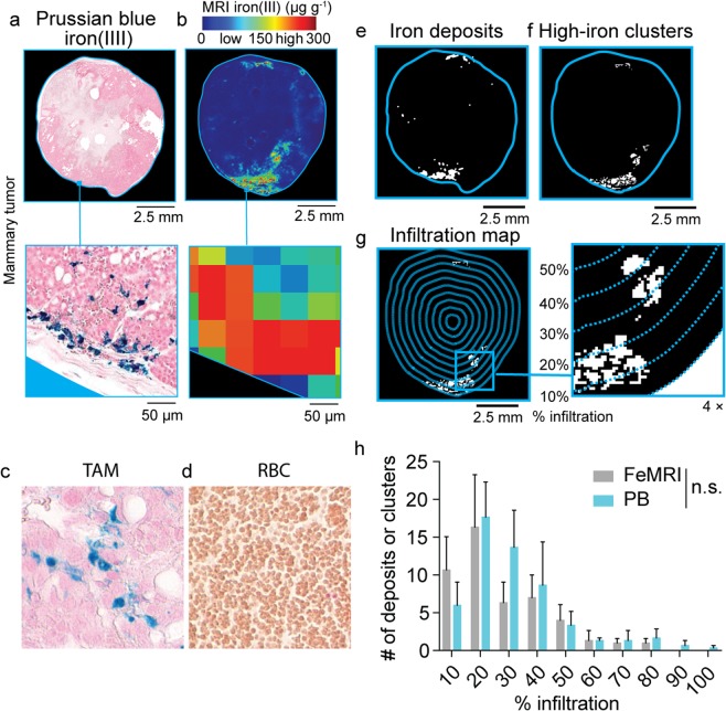
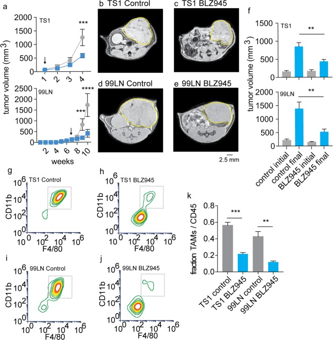
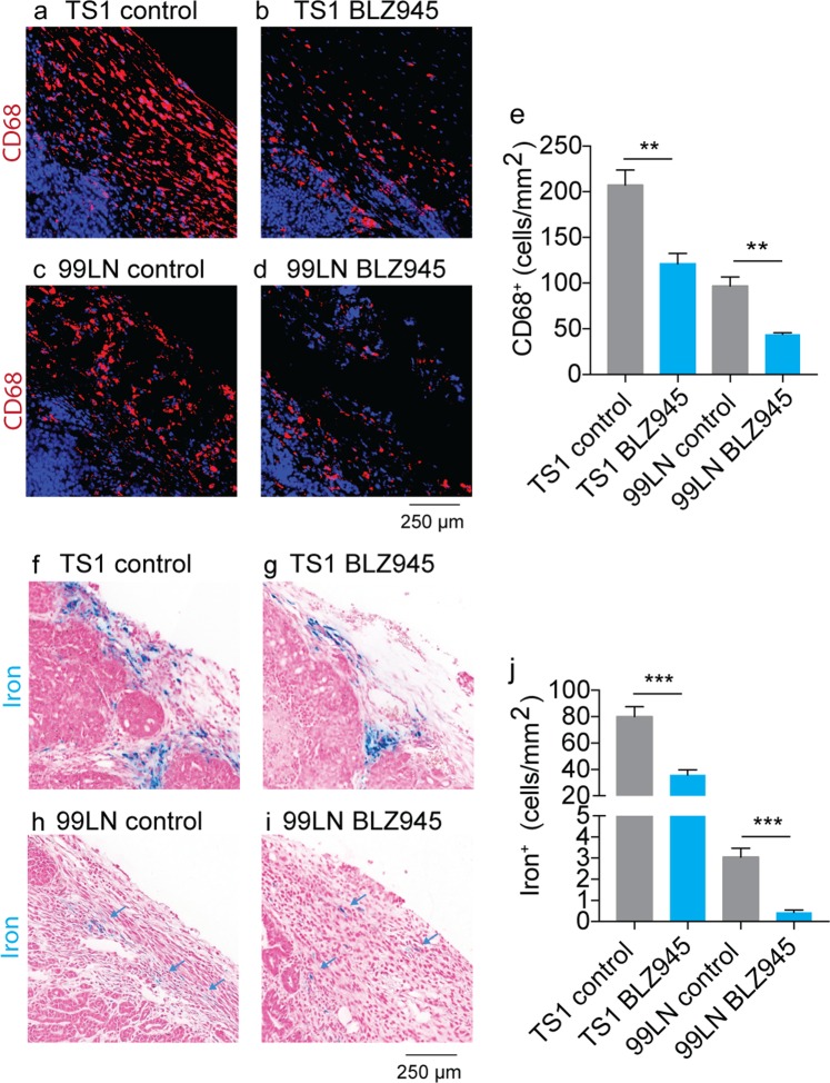
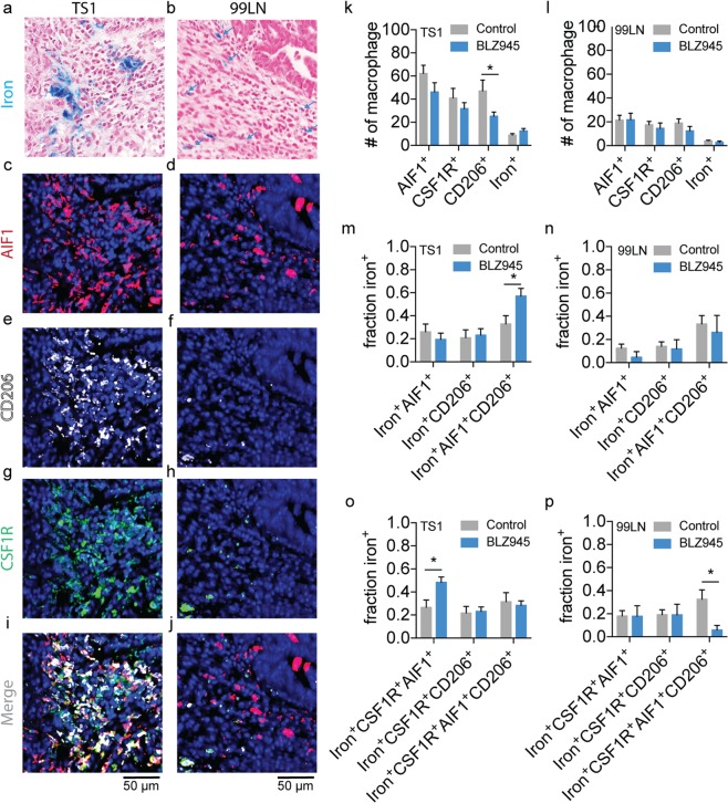
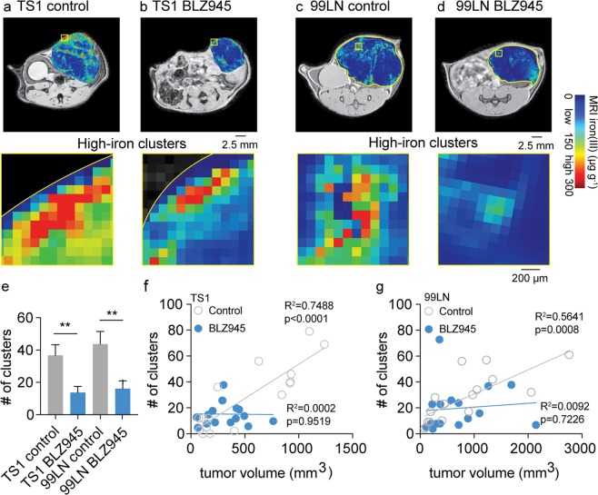
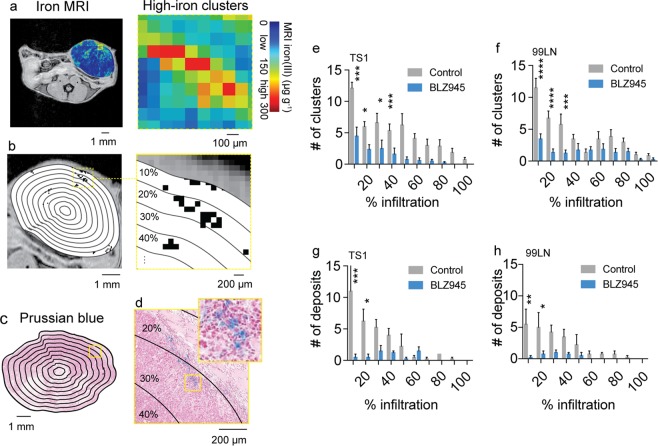
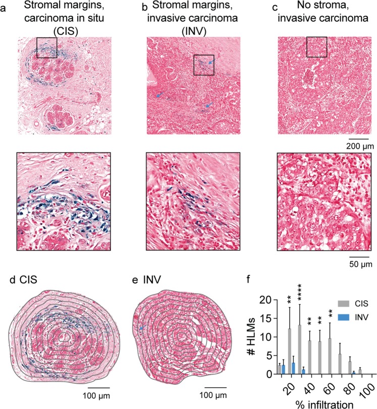
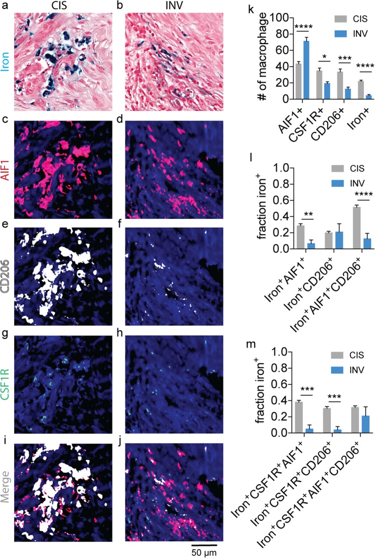
Iron deposits are a phenotypic trait of tumor-associated macrophages (TAMs). Histological iron imaging and contrast-agent free magnetic resonance imaging (MRI) can detect these deposits, but their presence in human cancer, and correlation with immunotherapeutic response is largely untested. Here, primarily using these iron imaging approaches, we evaluated the spatial distribution of polarized macrophage populations containing high endogenous levels of iron in preclinical murine models and human breast cancer, and used them as metabolic biomarkers to correlate TAM infiltration with response to immunotherapy in preclinical trials. Macrophage-targeted inhibition of the colony stimulating factor 1 receptor (CSF1R) by immunotherapy was confirmed to inhibit macrophage accumulation and slow mammary tumor growth in mouse models while also reducing hemosiderin iron-laden TAM accumulation as measured by both iron histology and in vivo iron MRI (FeMRI). Spatial profiling of TAM iron deposit infiltration defined regions of maximal accumulation and response to the CSF1R inhibitor, and revealed differences between microenvironments of human cancer according to levels of polarized macrophage iron accumulation in stromal margins. We therefore demonstrate that iron deposition serves as an endogenous metabolic imaging biomarker of TAM infiltration in breast cancer that has high translational potential for evaluation of immunotherapeutic response.


Conflict of interest statement

The authors declare no competing interests.

Figures

Figure 1
Figure 1
Imaging and spatial profiling of tumor macrophage iron deposits ex vivo with iron MRI (FeMRI) and Prussian blue iron histology. (a) Representative Prussian blue iron histology of MMTV-PyMT TS1 mammary tumor cross-section. Scale bar 2.5 mm. Expansion shows iron+ macrophage deposits. Expansion scale bar 50 μm. (b) Representative ex vivo FeMRI of MMTV-PyMT TS1 mammary tumor cross-section. Scale bar 2.5 mm. Expansion shows high-iron pixel clusters. Expansion scale bar 50 μm. (c) Representative Prussian Blue iron histology of tumor associated macrophages (TAM), and (d) red blood cells (RBC). Note iron+ macrophages and iron RBCs. (e) Iron deposit mask from Prussian blue histology. Scale bar 2.5 mm. (f) High-iron FeMRI cluster mask. Scale bar 2.5 mm. (g) Infiltration mapping using radial decile region rake sampling overlaid on high-iron MRI cluster mask. Scale bar 2.5 mm. 4x expansion shows high-iron FeMRI clusters and decile boundaries. (h) Infiltration profile showing number of histological iron deposits from Prussian blue (PB) and high-iron FeMRI pixel clusters (FeMRI) as a function of percent (%) infiltration into the MMTV-PyMT TS1 mammary tumors. (mean + s.e.m. n = 3 tissue cross-sections, n.s. p > 0.05 Kolmogorov-Smirnov test).
Figure 2
Figure 2
Validation of CSF1R immunotherapy effects on tumor growth and TAM accumulation in PyMT-MMTV breast cancer models. (a) Murine breast tumor models were established by orthotopic mammary fat pad injections and measured by caliper in TS1 and 99LN models during administration of the CSF1R inhibitor BLZ945 (200 mg/kg). Arrows indicate start of BLZ945 treatment (mean ± s.e.m. n = 5 mice/group, ***p < 0.001, ****p < 0.0001, 2-way ANOVA with Tukey’s multiple comparison test). Representative first-echo images from MGE MRI experiments made at study endpoints in control and BLZ945 treated (b,c) TS1 and (d,e) 99LN MMTV-PyMT models. (f) Pre-treatment and endpoint tumor volumes measured by MRI in the CSF1R inhibitor trials. (mean + s.e.m., n = 8 mice/group, **p < 0.01, two-tailed unpaired students t-test). Flow cytometry panels of TAMs (live CD45+Ly6cLy6g cells gated on CD11b+F4/80+ cells) obtained from control and BLZ945 treated (g,h) TS1, and (i,j) 99LN tumors. (k) TAM frequency with respect to total CD45+ myeloid cells in the TS1 and 99LN CSF1R inhibitor trials (mean + s.e.m. n = 4 mice/group, **p < 0.01, ***P < 0.001, two-tailed unpaired students t-test).
Figure 3
Figure 3
CD68 immunofluorescence and Prussian blue imaging of macrophage iron deposits in MMTV-PyMT murine breast cancer models of CSF1R immunotherapy. Representative CD68 macrophage immunofluoresent imaging in control and BLZ945 treated (a,b) TS1, and (c,d) 99LN tumors. Scale bar 250 µm (e) Absolute counts of CD68+ macrophages per mm2 MMTV-PyMT tumor cross-section in BLZ945 CSF1R inhibitor trials (mean + s.e.m. n = 4 mice/group, **p < 0.01, two-tailed unpaired students t-test). Representative iron staining using Prussian blue iron histochemistry in control and BLZ945 treated MMTV-PyMT (f,g) TS1, and (h,i) 99LN tumors. Scale bar 250 µm (j) Absolute counts of iron+ macrophages per mm2 tumor cross-section in the CSF1R inhibitor trials (mean + s.e.m. n = 4 mice/group, ***p < 0.001, two-tailed unpaired students t-test).
Figure 4
Figure 4
Immunofluorescent and Prussian blue imaging of macrophage iron deposit polarization and CSF1R status in MMTV-PyMT murine breast cancer models of CSF1R immunotherapy. Representative registered fields centered on TAM iron deposits in MMTV-PyMT TS1 and 99LN tumors stained for iron with Prussian blue (a,b), allograft inflammatory factor-1 (AIF1, M1-like, c,d), mannose receptor (CD206, M2-like, e,f), colony stimulating factor 1 receptor (CSF1R, g,h) and the combined immunofluorescent channels(i,j). Note fields of control tumors are shown, but are representative of both control and BLZ945 treated groups. Each field is 200 µm × 200 µm. Scale bar 50 µm. Blue arrows indicate location of iron+ macrophage in 99LN field. Number (#) of AIF1+, CSF1R+, CD206+ and iron+ macrophages detected per field for control and BLZ945 treated (k) TS1 and (l) 99LN MMTV-PyMT mammary tumor models (mean + s.e.m. n = 20 fields for TS1 control and BLZ945, n = 20 fields for 99LN control and n = 7 fields for 99LN BLZ945, *p < 0.05, Mann-Whitney test). Fraction of total iron+AIF1+, iron+CD206+, and iron+AIF1+CD206+ macrophages detected per field in control and BLZ945 treated groups for (m) TS1 and (n) 99LN mammary tumor models. Fraction of total iron+CSF1R+AIF1+, iron+CSF1R+CD206+ and iron+CSF1R+AIF1+CD206+ macrophages detected per field in control and BLZ945 treated groups for (o) TS1 and (p) 99LN mammary tumor models (mean + s.e.m. n = 20 fields for TS1 control, n = 20 fields TS1 BLZ945, n = 20 fields for 99LN control and n = 7 fields for 99LN BLZ945, *p < 0.05, Mann-Whitney test).
Figure 5
Figure 5
In vivo iron MRI (FeMRI) of murine macrophage iron deposits and correlation between immune and therapeutic CSF1R inhibitor response. Representative in vivo FeMRI axial cross sections of the mammary tumors are shown in control and BLZ945 treated (a,b) TS1, and (c,d) 99LN models. Scale bar 2.5 mm. Expansions show high-iron pixel clusters. Scale bar 200 µm. (e) Number (#) of high-iron FeMRI pixel clusters in the TS1 and 99LN tumors in the CSF1R inhibitor trials (mean + s.e.m. n = 8 mice/group, **p < 0.01 two-tailed unpaired students t-test). Linear correlations between high-iron FeMRI clusters and tumor volumes in the control(ο) and BLZ945-treated(•) (f) TS1 and (g) 99LN MMTV-PyMT tumor models (n = 8 mice/group, R2 and correlation p-value from linear Pearson correlation are shown).
Figure 6
Figure 6
Spatial scores of murine tumor macrophage infiltration in CSF1R immune therapy trials from iron MRI and Prussian blue iron histology. (a) In vivo iron MRI (FeMRI) of MMTV-PyMT mammary tumor. Scale bar 1 mm. Expansion shows high-iron pixel clusters. Scale bar 100 µm. (b) Binary high-iron pixel cluster mask from FeMRI of MMTV-PyMT tumor cross-section. Scale bar 1 mm. Concentric rake region of interest grid overlay used to profile clusters is shown. Expansion shows detected clusters and concentric counting grid. Scale bar 200 µm. (c) Prussian blue iron stained cross-section of MMTV-PyMT tumor showing the rake grid overlay used to score the iron deposits. Scale bar 1 mm. (d) Expansion showing deposit, i.e. colony of iron+ macrophages. Scale bar 200 µm. Infiltration profiles of high-iron clusters from FeMRI in control and BLZ945-treated (e) TS1 and (f) 99LN MMTV-PyMT models (mean + s.e.m. n = 8 mice/group, *p < 0.05, ***p < 0.001, ****p < 0.0001, 2-way ANOVA with Sidak’s multiple comparison test). Iron+ macrophage deposit infiltration profiles from Prussian blue histology in control and BLZ945-treated (g) TS1 and (h) 99LN MMTV-PyMT models (mean + s.e.m. n = 4 mice/group, *p < 0.05, ***p < 0.001, ****p < 0.0001, 2-way ANOVA with Sidak’s multiple comparison test).
Figure 7
Figure 7
Spatial scores of iron deposits from Prussian blue histology in human breast cancer. Prussian Blue iron histochemistry shows the presence of iron deposits in (a) stromal margins of carcinoma in situ (CIS) and (b) invasive carcinoma (INV, blue arrows). No iron deposits were associated with (c) invasive carcinoma exhibiting poorly defined stromal margins. Scale bar 200 µm. Expansions of boxes in (ac) shown below. Scale bar 40 µm. Concentric rake region of interest grid overlay used to profile HLMs in (d) CIS and (e) INV fields. Scale bar 100 µm. (f) Iron+ macrophage (HLM) infiltration profiles from Prussian blue histology in CIS and INV fields. (mean + s.e.m. n = 5 fields/cancer subtype, **p < 0.01, ****p < 0.0001, 2-way ANOVA with Sidak’s multiple comparison test).
Figure 8
Figure 8
Immunofluorescent and Prussian blue imaging of human macrophage iron deposit polarization and CSF1R status. Representative registered fields of human carcinoma in situ (CIS) and invasive carcinoma where iron deposits are found (INV) stained for iron with Prussian blue (a,b), allograft inflammatory factor-1 (AIF1, c,d), mannose receptor (CD206, e,f), colony stimulating factor 1 receptor (CSF1R, g,h) and the combined immunofluorescent channels (i,j). Each field is 200 µm × 200 µm. Scale bar 50 µm. (k) Number (#) of AIF1+, CSF1R+, CD206+ and iron+ macrophages detected per CIS and INV field. (mean+s.e.m. n=18 fields for CIS, n=9 fields for INV, *p < 0.05, ***p < 0.001, ****p < 0.0001, Mann-Whitney test). (l) Fraction of total iron+AIF1+, iron+CD206+, and iron+AIF1+CD206+ macrophages detected per CIS and INV field. (m) Fraction of total iron+CSF1R+AIF1+, iron+CSF1R+CD206+, and iron+CSF1R+AIF1+CD206+ macrophages detected per CIS and INV field (mean+s.e.m. n=18 fields for CIS, n=10 fields for INV, **p < 0.01, ***p < 0.001, ****p < 0.0001, Mann-Whitney test).

References

    1. Gentles AJ, et al. The prognostic landscape of genes and infiltrating immune cells across human cancers. Nat. Med. 2015;21:938–945. doi: 10.1038/nm.3909. - DOI - PMC - PubMed
    1. Leek RD, et al. Association of macrophage infiltration with angiogenesis and prognosis in invasive breast carcinoma. Cancer Res. 1996;56:4625–4629. - PubMed
    1. Pollard JW. Tumor-educated macrophages promote tumor progression and metastasis. Nat. Rev. Cancer. 2004;4:71–78. doi: 10.1038/nrc1256. - DOI - PubMed
    1. Quail DF, Joyce JA. Microenvironmental regulation of tumor progression and metastasis. Nature medicine. 2013;19:1423–1437. doi: 10.1038/nm.3394. - DOI - PMC - PubMed
    1. Gordon S, Pluddemann A, Estrada FM. Macrophage heterogeneity in tissues: phenotypic diversity and functions. Immunol. Rev. 2014;262:36–55. doi: 10.1111/imr.12223. - DOI - PMC - PubMed

Publication types

MeSH terms