. 2019 Jan 29;9(1):920.
doi: 10.1038/s41598-018-37000-z.

Impaired non-homologous end joining in human primary alveolar type II cells in emphysema

Affiliations

Impaired non-homologous end joining in human primary alveolar type II cells in emphysema

Beata Kosmider et al. Sci Rep. .

Abstract

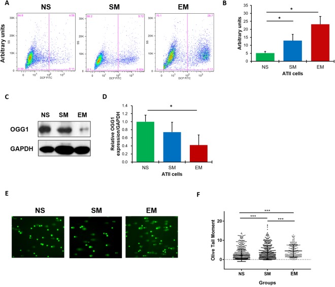
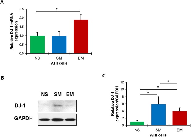
Emphysema is characterized by alveolar wall destruction induced mainly by cigarette smoke. Oxidative damage of DNA may contribute to the pathophysiology of this disease. We studied the impairment of the non-homologous end joining (NHEJ) repair pathway and DNA damage in alveolar type II (ATII) cells and emphysema development. We isolated primary ATII cells from control smokers, nonsmokers, and patients with emphysema to determine DNA damage and repair. We found higher reactive oxygen species generation and DNA damage in ATII cells obtained from individuals with this disease in comparison with controls. We also observed low phosphorylation of H2AX, which activates DSBs repair signaling, in emphysema. Our results indicate the impairement of NHEJ, as detected by low XLF expression. We also analyzed the role of DJ-1, which has a cytoprotective activity. We detected DJ-1 and XLF interaction in ATII cells in emphysema, which suggests the impairment of their function. Moreover, we found that DJ-1 KO mice are more susceptible to DNA damage induced by cigarette smoke. Our results suggest that oxidative DNA damage and ineffective the DSBs repair via the impaired NHEJ may contribute to ATII cell death in emphysema.


Conflict of interest statement

The authors declare no competing interests.

Figures

Figure 1
Figure 1
High ROS, DNA damage and the impairment of DNA damage repair in ATII cells in emphysema patients. ROS levels were analyzed in freshly isolated ATII cells from control non-smokers (NS), smokers (SM) and emphysema patients (EM) using DCF staining by flow cytometry. Representative images of flow cytometry profiles (A) and quantification (B) are shown. Representative Western blot images of OGG1 expression (C) and densitometric quantification of OGG1 expression normalized to GAPDH and non-smokers (D). Comet assay was used to detect DNA damage in freshly isolated ATII cells (E). Quantification of the Olive Tail Moment is also shown (F). Data are shown as mean values ± s.e.m. (N = 6 per group; *p < 0.05; ***p < 0.001, magnification 10 × 40).
Figure 2
Figure 2
High DNA damage and the impairment of DNA damage repair in ATII cells in patients with emphysema. Freshly isolated ATII cells from non-smokers (NS), smokers (SM) and patients with emphysema (EM) were used to determine γH2AX expression by Western blotting (A). Densitometric quantification of γH2AX levels normalized to GAPDH and non-smokers (B). Human lung sections were co-stained for SP-C to identify ATII cells (red), γH2AX (green) and nuclei (DAPI, blue) by immunohistofluorescence (magnification 10 × 63) (C). The relative fluorescence intensity of γH2AX expression in ATII cells is also shown (D). Representative p53 and p21 expression from the same Western blotting (E) and densitometric analysis (F) in ATII cells obtained from non-smokers, smokers and emphysema patients. Apoptotic ATII cells were detected using SP-C (red), caspase 3 (green) and DAPI (blue) staining by immunohistofluorescence (G) and quantification is also shown (H). Decreased number of ATII cells using SP-C staining (red) in lung tissue sections from emphysema patients by immunohistofluorescence (DAPI, blue; magnification 10 × 63) (I) and quantification (J). Data are presented as means ±s.e.m (*p < 0.05). Cultured ATII cells isolated from control non-smokers were treated with CSE for 24 h (K). Lane 1 – control, lane 2–1% CSE, lane 3–3% CSE and lane 4–6% CSE. Cell lysates were subjected to Western blotting to determine protein expression (N = 3 per group).
Figure 3
Figure 3
The impaired NHEJ DNA damage repair pathway in ATII cells and lung tissue in emphysema. 53BP1 expression in freshly isolated ATII cells from control non-smokers (NS), smokers (SM) and patients with emphysema (EM) was analyzed by Western blotting (A). Densitometric quantification is also shown (B). 53BP1 (green) expression is shown in ATII cells identified by proSP-C staining (red) in lung tissue sections (nuclei - DAPI, blue) by immunohistofluorescence (magnification 10 × 63) (C). Quantification of fluorescence intensity is also shown (D). DNA ligase IV (E,F) and XRCC4 (G,H) expression in freshly isolated ATII cells was determined by Western blotting and densitometric quantification. XLF levels in ATII cells were determined by Western blotting (I), densitometric quantification (J) and RT-PCR (K). XLF levels were analyzed in lung tissue obtained from control non-smokers and smokers, and areas with mild (ME) and severe emphysema (SE) from the same patient by Western blotting (L,M). Expression of OGG1 (N,O) was analyzed in lung tissue obtained from areas with mild and severe emphysema from the same patient by Western blotting and quantification. Representative Western blotting images and densitometric quantification are shown for PARP1 (P,Q), DNA ligase III (R,S) and RAD51 expression (T,U) in freshly isolated ATII cells. Data are shown as mean values ± s.e.m. (N = 7 per group; *p < 0.05).
Figure 4
Figure 4
DJ-1 expression in human ATII cells. DJ-1 expression was analyzed by RT-PCR (A), Western blotting (B) and densitometric analysis (C) in freshly isolated ATII cells from non-smokers (NS), smokers (SM) and patients with emphysema (EM). Data are shown as mean values ± s.e.m. (N = 6 per group; *p < 0.05).
Figure 5
Figure 5
DJ-1 and XLF interaction in freshly isolated ATII cells from patients with emphysema identified by mass spectrometry analysis. DJ-1 was co-immunoprecipitated in ATII cells obtained from non-smokers (NS), smokers (SM) and patients with emphysema (EM) followed by mass spectrometry analysis. The presence of DJ-1 (A) and its interaction with XLF (B) is shown in emphysema (N = 3 per group). Results were validated by co-immunoprecipitation of DJ-1 followed by Western blotting analysis of XLF expression in ATII cells obtained from non-smokers, smokers and patients with emphysema (C) and densitometric quantification (N = 6 per group; *P < 0.05) (D). Data is shown as means ± s.e.m.
Figure 6
Figure 6
XLF and DJ-1 expression in lung tissue obtained from mice exposed to cigarette smoke in vivo. Lung tissue was obtained from wild-type (WT) and DJ-1 KO mice exposed to 150 mg/m3 TSP as described in Material and Method section (CTL – control; CS-cigarette smoke). The absence of DJ-1 expression in DJ-1 KO mice was confirmed by Western blotting (A). Representative Western blot images of expression and densitometric analysis of XLF (B,C), γH2AX (D,E), p21 (F,G), 4-HNE (H,I) and caspase 3 (J,K) are shown. Densitometric quantification of protein expression was normalized to β-actin (N = 3 per group). *P < 0.05; **P < 0.001. Data is shown as means ± s.e.m.

References

    1. Barnes PJ. Role of HDAC2 in the pathophysiology of COPD. Annu Rev Physiol. 2009;71:451–464. doi: 10.1146/annurev.physiol.010908.163257. - DOI - PubMed
    1. Lokke A, Lange P, Scharling H, Fabricius P, Vestbo J. Developing COPD: a 25 year follow up study of the general population. Thorax. 2006;61:935–939. doi: 10.1136/thx.2006.062802. - DOI - PMC - PubMed
    1. Gogebakan B, et al. The role of bronchial epithelial cell apoptosis in the pathogenesis of COPD. Mol Biol Rep. 2014;41:5321–5327. doi: 10.1007/s11033-014-3403-3. - DOI - PubMed
    1. Abboud RT, Vimalanathan S. Pathogenesis of COPD. Part I. The role of protease-antiprotease imbalance in emphysema. Int J Tuberc Lung Dis. 2008;12:361–367. - PubMed
    1. Baluchamy S, et al. Induction of cell death through alteration of oxidants and antioxidants in lung epithelial cells exposed to high energy protons. J Biol Chem. 2010;285:24769–24774. doi: 10.1074/jbc.M110.138099. - DOI - PMC - PubMed

Publication types